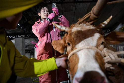
Peritonitis, or inflammation of the peritoneal cavity, is a serious and often life-threatening condition in cattle. It typically results from gastrointestinal tract perforations, uterine rupture, abdominal trauma, or post-surgical complications such as those following cesarean sections or abomasopexy. Diagnosing peritonitis in cattle can be challenging, particularly in the early stages when clinical signs are subtle. Traditional diagnostic methods such as clinical examination, hematology, or exploratory laparotomy are either non-specific, invasive, or impractical in field conditions.

Ultrasound-guided fluid aspiration has emerged as a non-invasive and effective diagnostic technique that allows for both visual evaluation of the abdominal cavity and sample collection for cytological and bacteriological analysis. This method is gaining popularity in veterinary practice worldwide, particularly in North America and Europe, due to its high diagnostic accuracy and minimal risk to the animal. This article explores the diagnostic value of ultrasound-guided fluid aspiration in cattle with suspected peritonitis, drawing from international veterinary practices and field experience.
Understanding Peritonitis in Cattle
Peritonitis in cattle can be localized or generalized, acute or chronic. It is typically associated with infections caused by Escherichia coli, Trueperella pyogenes, or other mixed anaerobic and aerobic bacteria. The primary clinical signs include:
Fever
Abdominal distension
Pain on palpation
Reduced rumen motility
Decreased appetite and milk production
Tachycardia and dehydration
However, these symptoms are not specific and can overlap with other abdominal disorders such as displaced abomasum, traumatic reticuloperitonitis (hardware disease), or intestinal volvulus. Thus, accurate diagnosis becomes crucial for timely and effective treatment.
Role of Ultrasound in Diagnosing Peritonitis
Ultrasound imaging has transformed the way veterinarians approach abdominal diseases in cattle. B-mode ultrasonography allows for real-time, non-invasive visualization of internal structures, detection of abnormal fluid accumulation, fibrin deposits, abscesses, and organ displacement. It has become a critical diagnostic tool in both clinical and field settings.
In suspected peritonitis cases, the veterinarian typically uses a low-frequency (2.5–5.0 MHz) convex or microconvex probe to examine the ventral and lateral abdomen. Signs suggesting peritonitis on ultrasound may include:
Anechoic or echogenic fluid within the abdominal cavity
Fibrinous strands floating within the fluid
Loculated abscesses or pockets of gas
Thickened mesenteric or peritoneal layers
While these findings are indicative, they are not definitive. Thus, coupling ultrasonographic imaging with fluid aspiration for laboratory evaluation provides a more conclusive diagnosis.
Ultrasound-Guided Fluid Aspiration: Technique and Application
Ultrasound-guided abdominocentesis involves inserting a needle into a fluid pocket under real-time ultrasound guidance to collect peritoneal fluid. This approach offers several advantages:
Precision: Ensures accurate placement of the needle, avoiding organs and minimizing trauma.
Sample Quality: Improves chances of retrieving diagnostic-quality fluid.
Safety: Reduces risk of iatrogenic injury and infection.
Procedure:
Site Selection: The most common site for fluid aspiration is the right or left ventral abdominal region, between the umbilicus and the udder or prepuce.
Preparation: The area is clipped, scrubbed, and sterilized. Local anesthesia may be applied.
Probe Guidance: The ultrasound probe identifies a fluid pocket. The needle is advanced under continuous visualization.
Fluid Collection: Once fluid is aspirated, it is submitted for cytology, culture, and sensitivity testing.
In field conditions, portable ultrasound devices like the BXL-V50 have proven to be highly practical due to their long battery life, waterproof design, and real-time imaging capabilities.
Interpretation of Peritoneal Fluid
The appearance and composition of the aspirated fluid help confirm the presence and type of peritonitis:
Normal fluid is scant and straw-colored with low protein and cell count.
Inflammatory exudate is turbid or bloody, with elevated protein (>3 g/dL) and high nucleated cell counts (>10,000 cells/μL).
Septic peritonitis often yields purulent, malodorous fluid with degenerative neutrophils and intracellular bacteria on cytology.
The presence of fibrin, bacteria, and elevated lactate concentrations are strong indicators of septic processes requiring immediate treatment. Gram staining and culture provide information about the causative agents, guiding antibiotic therapy.
Advantages Over Traditional Methods
1. Improved Accuracy and Early Detection:
Ultrasound can detect fluid pockets too small to identify on physical exam or via blind abdominocentesis. This improves the chances of early diagnosis and treatment before the condition becomes irreversible.
2. Minimally Invasive:
Compared to laparotomy or exploratory surgery, ultrasound-guided aspiration requires only a needle puncture, reducing animal stress and recovery time.
3. Real-Time Decision Making:
Veterinarians can immediately assess whether further action is needed (e.g., surgery, euthanasia, or aggressive medical treatment) based on fluid characteristics.
4. Field-Friendly:
With advancements in portable ultrasound systems like the BXL-V50, field veterinarians can perform this diagnostic procedure even in remote locations without requiring referral to a veterinary hospital.

Limitations and Considerations
Despite its many benefits, this technique is not without limitations:
False negatives may occur if fluid is loculated or if the disease is localized without significant fluid accumulation.
Operator dependence: Skill and experience in both ultrasound imaging and fluid interpretation significantly affect diagnostic accuracy.
Cost and equipment availability: Although portable ultrasound units have become more affordable, cost may still be a barrier in some regions.
Moreover, even when fluid is collected, a definitive diagnosis often requires integration of clinical signs, ultrasonographic findings, and laboratory results.
Global Perspective and Adoption
In North America and Europe, ultrasound-guided fluid aspiration is now widely practiced in university veterinary hospitals and increasingly adopted in commercial dairy and beef operations. Many veterinary schools incorporate this technique into training due to its safety, repeatability, and educational value.
A study published in the Journal of Dairy Science in 2022 reported that ultrasound-guided aspiration increased the diagnostic rate of peritonitis by over 35% compared to traditional methods. Additionally, a 2023 survey of European bovine practitioners revealed that over 60% of field veterinarians preferred this technique when evaluating cows with suspected abdominal infection.
In countries like Australia and New Zealand, where large-scale cattle farming is common, veterinarians favor this method for its ability to rapidly assess animals in open-range conditions without needing hospitalization.
Conclusion
Ultrasound-guided fluid aspiration has revolutionized the diagnosis of peritonitis in cattle. By offering a fast, non-invasive, and accurate assessment of the abdominal cavity, it provides veterinarians with valuable diagnostic and therapeutic insights. Coupled with appropriate laboratory analysis, this technique facilitates early intervention, improving recovery rates and reducing the economic impact of delayed or missed diagnoses.
As equipment becomes more portable and affordable—like the BXL-V50—this method is expected to become a standard diagnostic practice in both developed and developing agricultural systems. For any veterinarian managing cattle with signs of abdominal distress, mastering this technique can significantly enhance diagnostic confidence and improve patient outcomes.
References
LeBlanc, S. J., Lissemore, K. D., Kelton, D. F., Duffield, T. F., & Leslie, K. E. (2022). The diagnostic value of ultrasound-guided abdominocentesis in dairy cattle with peritonitis. Journal of Dairy Science, 105(3), 1740–1752.
Braun, U. (2009). Ultrasonography in gastrointestinal disease in cattle. Veterinary Clinics of North America: Food Animal Practice, 25(3), 567–590.
Peek, S. F., & Divers, T. J. (2018). Rebhun's Diseases of Dairy Cattle (3rd ed.). Saunders.
Beef Cattle Institute (2023). “Use of Ultrasound for Growth Evaluation in Cattle.”
tags:
Text link:https://www.bxlultrasound.com/ns/858.html


