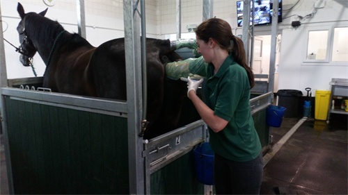
In modern equine reproductive medicine, ensuring the health and fertility of broodmares is crucial for breeding programs worldwide. One of the most common and significant reproductive issues in mares is the accumulation of intrauterine fluid, often a sign of inflammation, poor uterine clearance, or underlying anatomical abnormalities. Fortunately, real-time ultrasonography offers an effective, non-invasive method for detecting and monitoring such abnormalities, providing veterinarians and horse breeders with critical insights to guide treatment and management.

This article explores how equine reproductive tract abnormalities—especially uterine fluid retention—can be accurately diagnosed through ultrasound, and how real-time imaging has revolutionized reproductive care for horses globally.
Understanding the Equine Reproductive Tract
The mare's reproductive tract consists primarily of the ovaries, oviducts, uterus, cervix, and vagina. The uterus is a key structure in pregnancy and must maintain a sterile, fluid-free environment for successful conception and gestation. However, due to anatomical or physiological dysfunctions—often worsened with age, multiple pregnancies, or infection—the uterus may fail to clear inflammatory secretions or post-mating fluid effectively. This leads to the abnormal retention of fluid, commonly associated with endometritis or delayed uterine clearance.
Mares with this condition may appear healthy externally, showing no clear behavioral signs of distress, yet struggle with conception or repeated early embryonic loss. That’s where real-time ultrasound becomes essential.
The Role of Real-Time Ultrasound in Uterine Fluid Detection
Real-time B-mode ultrasound allows clinicians to visualize the internal reproductive structures of the mare in motion. Through transrectal scanning, the uterus can be imaged to assess wall thickness, luminal contents, and any signs of fluid accumulation. Fluid pockets appear as anechoic (dark) areas on the ultrasound screen and may vary in volume, distribution, and echogenicity depending on the cause.
Why is this important?
Detecting even small amounts of intrauterine fluid early can drastically alter the clinical outcome. According to equine reproductive specialists in the United States and Europe, ultrasonography is now a first-line tool for diagnosing uterine pathology, particularly in mares with breeding failures. Early intervention—whether through uterine lavage, oxytocin therapy, or antibiotics—depends on accurate, real-time diagnosis.
When and How to Scan
Ultrasound examination of the mare’s reproductive tract is typically performed during key stages:
Before breeding: To ensure the uterus is clear of fluid and to evaluate follicular development.
Post-breeding (24–48 hours): To assess post-mating endometritis and check for retained fluid.
During early pregnancy: To confirm conception and monitor embryonic viability.
In mares with history of infertility: For ongoing monitoring of uterine health.
In practice, veterinarians insert a lubricated transrectal probe—such as those compatible with the BXL-V50 portable ultrasound system—to obtain high-resolution images of the uterus. The BXL-V50, commonly used in veterinary clinics across Europe and North America, provides clear visualization of uterine abnormalities, including fluid presence, cysts, or fibrotic changes. Its real-time imaging and waterproof design make it ideal for equine use in various environments.
Clinical Presentation of Uterine Fluid
Ultrasound imaging helps categorize uterine fluid abnormalities:
Simple Anechoic Fluid: Often a post-ovulatory or post-mating accumulation, which typically resolves with oxytocin.
Echogenic or Flocculent Fluid: Indicates inflammatory cells or pus; may suggest acute or chronic endometritis.
Localized Fluid Pockets: Sometimes linked to anatomical abnormalities like uterine cysts or fibrotic scarring.
Free-floating Debris: May suggest chronic inflammation or contamination during breeding.
Veterinarians often measure the volume and distribution of fluid and use follow-up scans to assess treatment response. If fluid persists, additional diagnostics—like cytology or uterine biopsy—may be performed.

Risk Factors and Prevalence
Research from the University of Kentucky’s Gluck Equine Research Center indicates that up to 15% of mares show signs of delayed uterine clearance post-mating, with the risk increasing in older or multiparous mares. Anatomical issues, such as a pendulous uterus, tight cervix, or poor uterine tone, exacerbate the problem.
In the UK, studies show that uterine fluid detected via ultrasound in pre-breeding exams correlates with a 40–60% drop in pregnancy rates if left untreated. These statistics emphasize the importance of early, routine ultrasound assessments for mares, especially those with a history of infertility.
Management Strategies Based on Ultrasound Findings
Once fluid is detected, real-time ultrasonography continues to play a role in monitoring the mare’s response to treatment. Common therapeutic options include:
Uterine lavage: Physically flushing out fluid and debris with sterile saline.
Oxytocin injections: Stimulate uterine contractions to expel residual fluid.
Systemic or intrauterine antibiotics: For bacterial endometritis, often guided by culture and sensitivity testing.
Prostaglandins: Used in some cases to induce estrus and facilitate uterine clearance.
Veterinarians in the U.S. and Australia emphasize a “scan-treat-rescan” protocol, using ultrasound to guide decision-making every step of the way.
Ultrasound Beyond Fluid Detection
While detecting intrauterine fluid is a primary function, ultrasonography in mares also assists in:
Diagnosing ovarian abnormalities (e.g., anovulatory follicles or granulosa cell tumors)
Identifying uterine cysts: Which can mimic early embryos or interfere with implantation
Monitoring pregnancy progression: Including fetal heartbeat and movement
Guiding artificial insemination timing: By tracking follicular growth and ovulation
Many equine reproduction experts advocate for routine use of ultrasound throughout the breeding season, not just in problem mares.
International Perspectives on Equine Reproductive Ultrasound
Across the globe, the veterinary consensus is clear: real-time ultrasonography is indispensable in equine reproductive management. In Germany, veterinary training includes detailed modules on ultrasound-guided mare care. In Canada and the U.S., portable ultrasound devices are standard in field practice. Countries like New Zealand and the Netherlands are exploring tele-ultrasound services, where images can be reviewed remotely by reproductive specialists.
Veterinarians also report growing interest in using ultrasound in embryo transfer programs, particularly to assess uterine receptivity and synchronization between donor and recipient mares.
Advantages of Real-Time Ultrasound in Mare Reproductive Health
✅ Non-invasive and low-stress: Mares tolerate rectal scans well with minimal sedation.
✅ High diagnostic accuracy: Real-time visualization eliminates guesswork.
✅ Rapid decision-making: Immediate results enable quick treatment initiation.
✅ Reproductive planning: Supports optimal breeding and embryo transfer timing.
✅ Cost-effective: Prevents unnecessary breeding attempts and reduces open mare days.
Conclusion
Detecting and managing reproductive tract abnormalities in mares is essential for the success of any breeding program. Among these, uterine fluid accumulation is one of the most common yet manageable conditions—if caught early. Real-time ultrasound has become the gold standard in equine gynecology, empowering veterinarians and breeders to identify issues, monitor treatments, and improve conception rates.
With advanced portable systems like the BXL-V50 and increased access to veterinary training worldwide, the integration of ultrasonography into routine mare care is not just a recommendation—it’s a necessity. As the equine industry continues to evolve, those who embrace real-time reproductive imaging will be better equipped to achieve sustainable breeding success.
Reference Sources:
McCue, P. M., & Ferris, R. A. (2021). Reproductive Ultrasonography in the Mare. Colorado State University Equine Reproduction Laboratory.
Riddle, W. T., & LeBlanc, M. M. (2020). Diagnosis and Treatment of Uterine Fluid Accumulation in Mares. American Association of Equine Practitioners.
Gluck Equine Research Center. (2022). Reproductive Performance and Uterine Health in Broodmares.
USDA-APHIS. (2019). Equine Health Monitoring and Reproductive Trends in the U.S.
tags:
Text link:https://www.bxlultrasound.com/ns/857.html


