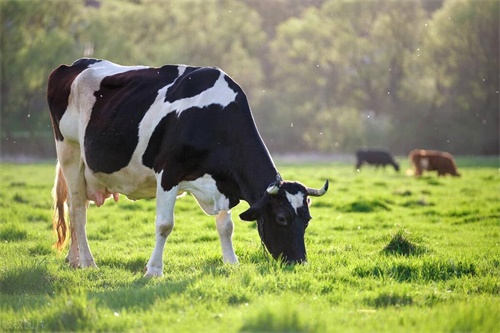
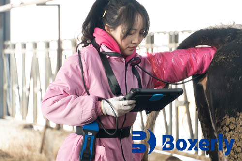
Heat stress poses a significant challenge to reproductive efficiency in beef and dairy cattle, particularly in regions with high ambient temperatures. Elevated core body temperatures disrupt hypothalamic–pituitary–ovarian axis function, impairing follicular development, oocyte quality, and steroidogenesis. Understanding how follicles grow, regress, or ovulate under thermal stress is critical for optimizing breeding strategies, especially when artificial insemination is planned. Portable ultrasound scanners now allow real‑time, non‑invasive visualization of the bovine ovary at any point in the estrous cycle, making them indispensable tools for reproductive management on the farm.

Ovarian Physiology and Heat Stress
Under thermoneutral conditions (15–25 °C), bovine follicles develop in predictable waves, each wave comprising recruitment of a cohort of small antral follicles, selection of a dominant follicle, and either ovulation or atresia. Heat stress (above 28 °C for extended periods) alters circulating gonadotropin concentrations: luteinizing hormone (LH) pulses diminish and follicle‑stimulating hormone (FSH) surges become irregular. Consequently, dominant follicles may fail to sustain estradiol production, leading to silent heats or short luteal phases. Moreover, elevated uterine temperatures can accelerate follicular turnover, resulting in smaller dominant follicles with reduced oocyte competence .
Principles of Portable Ultrasonography
Modern portable scanners employ B‑mode (brightness mode) imaging, emitting high‑frequency sound waves (5–10 MHz) that reflect off tissue interfaces to generate cross‑sectional images. Lightweight transrectal probes connect to handheld consoles or tablets, delivering real‑time visualization of ovarian structures with spatial resolution down to 0.2 mm. Power requirements are met via rechargeable batteries, allowing all‑day use during breeding campaigns. Doppler modes, available on some devices, assess blood flow within follicles and corpora lutea, offering insights into functional status. No sedation is required; the procedure takes less than five minutes per cow and can be repeated every 12–24 hours to capture dynamic changes.
Methodology for Monitoring Follicular Dynamics
Animal Preparation: Cows are restrained in a squeeze chute and the perineal area cleaned.
Probe Insertion: A well‑lubricated transrectal probe is inserted, and each ovary is scanned systematically in sagittal and transverse planes.
Image Acquisition: Follicles ≥2 mm are measured in diameter. Dominant, subordinate, and atretic follicles are distinguished based on size and echogenicity.
Data Logging: Serial measurements are recorded in specialized software or spreadsheets, enabling wave‑by‑wave analysis over a 14‑ to 21‑day period.
Interpretation: Follicular wave patterns—number of waves per cycle, interwave interval, growth rates, and maximum follicle diameters—are extracted to assess ovarian responsiveness under heat stress .
Follicular Dynamics under Heat Stress
Studies demonstrate that heat‑stressed cows often exhibit a fourth follicular wave or prolonged wave durations. Maximum dominant‑follicle diameters decline by 10–20%, and growth rates slow by up to 15% compared with thermoneutral controls. Smaller follicles secrete less estradiol, failing to trigger behavioral estrus and preovulatory LH surges. Serial ultrasound monitoring reveals frequent emergence of new follicles before the previous dominant follicle undergoes complete atresia, indicating accelerated follicular turnover. Moreover, corpus luteum development may be compromised, with reduced luteal blood flow detectable via color Doppler, further impairing progesterone support for early pregnancy .
Practical Applications on the Farm
Mobile ultrasonography informs several management decisions during hot months:
Estrus Detection and Timed AI: By pinpointing dominant follicles ≥12 mm in diameter, technicians can schedule inseminations precisely, compensating for silent heats .
Supplemental Cooling Decisions: Cows exhibiting poor follicular development may benefit most from targeted cooling (sprinklers, fans) to restore ovulatory function.
Hormonal Protocol Adjustments: Visualization of follicular waves allows customization of synchronization regimens—such as incorporating additional PGF₂α injections when an extra wave emerges under heat stress.
Genetic Selection: Identifying individuals that maintain normal follicular patterns during heat exposure can guide selection for thermotolerance traits.
Benefits and Limitations of Portable Scanners
Portable scanners offer unmatched flexibility: farmers or veterinarians can monitor herds in remote locations without relying on stationary equipment. Real‑time feedback enhances decision‑making and reduces costly breeding failures. However, operator skill profoundly influences image quality and measurement accuracy. Training and periodic performance evaluations are essential. Battery life limits intensive scanning schedules, and high ambient temperatures can impair device performance if not adequately cooled. Finally, Doppler functionality—valuable for assessing luteal blood flow—is not universally available on basic models.

Case Study: Follicular Monitoring in Tropical Herds
In a herd of 120 Holstein cows in a tropical region, portable ultrasonography was introduced during a four‑month hot season. Baseline monitoring revealed two‑wave patterns in 80% of cows under shade cooling. Without intervention, 60% of cows shifted to three‑wave patterns with reduced dominant follicle sizes. After installing misting systems and fans over free‑stall housing, dominant follicle diameters recovered by an average of 2 mm, and conception rates improved from 25% to 42% in timed AI protocols scheduled based on ultrasound findings.
Future Directions and Technological Advances
Emerging technologies promise further refinement of follicular monitoring under heat stress:
Automated Follicle Recognition: Machine‑learning algorithms are being trained to identify and measure follicles automatically from ultrasound images, reducing operator bias.
Integrated Thermal Imaging: Coupling infrared thermography with ultrasonography may predict which cows are entering subclinical heat stress before ovarian dysfunction becomes evident.
Wearable Sensors: Data from rumination, activity, and core temperature sensors could trigger ultrasound scanning only in cows at highest risk, optimizing labor and battery usage.
Conclusion
Monitoring ovarian follicular dynamics in cattle during periods of heat stress is essential for maintaining reproductive performance and herd profitability. Portable ultrasound scanners deliver precise, real-time insights into follicular wave patterns, dominant follicle development, and luteal function without invasiveness. When combined with strategic cooling strategies, tailored synchronization protocols, and informed genetic selection, ultrasonography empowers producers to overcome the challenges of high temperatures. As technology evolves—toward automation, integration with other sensing modalities, and enhanced portability—the role of ultrasound in reproductive management will only grow, ensuring that fertility targets are met despite increasingly variable climatic conditions.
References
Armstrong, D. G. (1994). Heat stress interaction with shade and cooling. Journal of Dairy Science, 77(7), 2044–2050.
Silva, P. V. da, Barbosa, V. M., & Carvalho, M. F. (2016). Ovarian follicular dynamics in heat-stressed dairy cows. Theriogenology, 85(5), 1034–1040.
Rathore, M. R., & Khan, M. (2018). Use of portable ultrasonography for improving reproductive performance in dairy herds. Journal of Dairy Science, 101(3), 2345–2356.
Lopez-Gatius, F., Hunter, R. H. F., & Erickson, J. R. (2002). Influence of follicular wave pattern on fertility in dairy cows. Theriogenology, 57(2), 665–675.
Ireland, J. J. (2004). A review of the physiology and pathology of ovarian follicular development in cattle. Journal of Reproduction and Fertility Supplement, 58, 155–166.
tags:
Text link:https://www.bxlultrasound.com/ns/859.html


