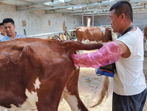
As a dairy farmer, maintaining the reproductive health of cows is one of the most critical factors in ensuring a profitable and sustainable operation. Reproductive efficiency directly affects milk production, calving intervals, and long-term herd productivity. Among the many technologies available today, real-time ultrasonography—commonly referred to as animal ultrasound scanning—has emerged as a reliable, non-invasive, and highly informative tool for monitoring uterine health in dairy cows. In this article, I will explore how we utilize real-time ultrasound to evaluate and protect the uterine health of our dairy herd, and why this technology has become indispensable on many modern farms around the world.

Understanding Uterine Health in Dairy Cows
In dairy cows, the uterus plays a central role in successful reproduction. After calving, the uterus must undergo involution—a natural process where it returns to its pre-pregnancy size and condition. This process needs to occur efficiently for the cow to resume normal reproductive cycles and become pregnant again within a timely interval. Delays or complications in uterine recovery, such as retained placenta, metritis, or endometritis, can significantly reduce reproductive performance.
Foreign studies and dairy experts emphasize that early detection of uterine disorders is key to minimizing economic losses. This is where real-time ultrasound scanning has revolutionized veterinary reproductive management.
The Role of Real-Time Ultrasound in Uterine Health Evaluation
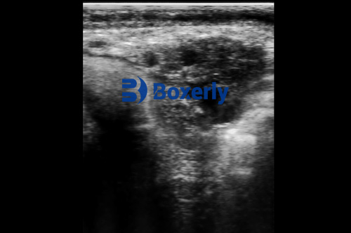
Real-time ultrasound allows us to visualize the internal structures of the uterus with great clarity. By applying a transrectal probe, veterinarians or trained farm staff can observe the uterine horns, body, and surrounding tissues in real-time, detecting fluid accumulation, tissue abnormalities, or inflammation.
Compared to traditional rectal palpation, which relies solely on tactile assessment, ultrasonography provides visual confirmation of uterine status. In foreign veterinary practice, especially across Europe and North America, real-time ultrasound has become the gold standard for early diagnosis of postpartum uterine disorders.
Common Uterine Conditions Identified by Ultrasound:
Uterine Involution Delay: Incomplete reduction in uterine size post-calving.
Metritis: Inflammation of the uterine wall, often characterized by fluid accumulation and echogenic debris.
Endometritis: Chronic inflammation presenting as irregularities in the uterine lining.
Retained Placenta Fragments: Visual identification of retained tissues that might not be detected manually.
Uterine Fluid Accumulation: Pockets of hypoechoic (dark) fluid that signal possible infection.

Advantages of Real-Time Ultrasonography on the Farm
Incorporating ultrasound scanning into routine reproductive management offers multiple advantages that foreign dairy producers have widely recognized:
1. Non-invasive and Stress-Free
Ultrasound scanning is non-invasive, causing minimal discomfort to the animal when performed correctly. This allows for frequent monitoring without compromising animal welfare.
2. Real-Time Results
Unlike laboratory tests that require sample collection and processing time, ultrasound provides immediate visual feedback, allowing for quick decision-making on treatment or management adjustments.
3. High Accuracy
Ultrasound delivers a high level of diagnostic accuracy, especially in detecting subclinical infections or early pathological changes that are difficult to identify by palpation alone.
4. Early Intervention
Early detection of uterine abnormalities enables prompt treatment, reducing the likelihood of chronic conditions and preserving reproductive performance.
5. Improved Fertility Outcomes
By closely monitoring uterine health, farmers can optimize breeding timing and improve conception rates, which ultimately leads to higher milk production and profitability.
Real-Life Application: My Experience Using Ultrasound for Uterine Health
On my farm, we incorporate real-time ultrasound as part of our postpartum cow monitoring program. Within two weeks post-calving, each cow undergoes a routine ultrasound exam to ensure proper uterine involution.
In many cases, the uterus returns to normal size without complications. However, when abnormalities are detected—such as retained fluid or tissue debris—we can initiate appropriate treatments such as antibiotics, uterine lavage, or hormonal therapies much earlier than we could by palpation alone.
One notable case involved a cow that showed no external symptoms of distress after calving. Upon routine ultrasound scanning, however, we identified hyperechoic material suggesting retained fetal membranes. Early intervention prevented the development of metritis and allowed the cow to resume her reproductive cycle without issue. Without ultrasound, this subclinical issue might have gone unnoticed until severe complications developed.

Real-Time Monitoring Throughout the Breeding Cycle
Ultrasound isn’t limited to postpartum care; it plays a vital role throughout the entire reproductive cycle:
Pre-Breeding Exams
Before artificial insemination, ultrasound can confirm normal uterine tone and detect ovarian structures like follicles and corpus luteum, ensuring the cow is at the optimal stage for breeding.
Pregnancy Diagnosis
From as early as 28 days post-breeding, ultrasound can confirm pregnancy, detect twin pregnancies, and identify embryonic viability—offering much earlier and more accurate information compared to palpation.
Monitoring Embryonic Development
In some advanced programs, ultrasound allows us to track embryo development, monitor for abnormalities, and make informed decisions about embryo transfer candidates.
Key Ultrasonographic Parameters for Uterine Assessment
During real-time scanning, several parameters are evaluated:
Uterine Wall Thickness: Increased thickness may indicate inflammation or infection.
Echogenicity of Uterine Contents: The appearance of the fluid (clear or cloudy) provides clues about infection status.
Size and Symmetry of Uterine Horns: Asymmetry may suggest involution delay or pathology.
Presence of Retained Tissues: Visualization of echogenic fragments points to incomplete expulsion of fetal membranes.
The Global Perspective: Adoption of Ultrasound in Dairy Farms Worldwide
Foreign literature and case studies highlight how real-time ultrasound has transformed dairy management internationally. In countries like the United States, Canada, the Netherlands, and Australia, ultrasound is now standard practice for both large-scale commercial dairies and smaller family-run operations.
According to the Journal of Dairy Science, farms incorporating routine ultrasound monitoring experience:
Shorter calving intervals
Reduced incidence of chronic uterine disease
Improved first-service conception rates
Decreased culling due to reproductive failure
These improvements translate into significant economic benefits, particularly when milk prices fluctuate and reproductive efficiency becomes the key to financial sustainability.

Economic Impact of Timely Uterine Health Monitoring
Timely monitoring of uterine health via ultrasound translates directly into farm profitability. Foreign economic models estimate that each day of delayed conception can cost a dairy farm between $2 and $5 per cow per day due to lost milk production and extended calving intervals.
When uterine disorders are left undetected, treatment costs increase, fertility declines, and in severe cases, cows may require early culling—leading to even greater financial losses.
By contrast, early diagnosis through ultrasound reduces veterinary costs, shortens treatment duration, and preserves valuable genetics within the herd.
Training and Implementation: Making Ultrasound Accessible to Farmers
One common misconception is that ultrasound requires extensive veterinary expertise. While professional veterinarians play a critical role, many foreign training programs now offer courses for farmers and farm managers, enabling them to conduct routine scans under veterinary supervision.
Portable, rugged ultrasound machines—such as the BXL-V50 Veterinary Ultrasound Scanner—are specifically designed for farm use. With features like waterproof housing, long battery life, high-definition imaging, and intuitive controls, devices like these make it feasible for even small farms to adopt real-time uterine monitoring.
In my own experience, the portability and durability of such devices have made daily scanning practical even under tough field conditions, regardless of weather or barn environment.

Limitations and Challenges
While ultrasound is a powerful tool, it is not without limitations:
Operator Dependency: Accuracy depends heavily on operator skill and experience.
Initial Cost: High-quality ultrasound equipment represents a significant initial investment.
Interpretation Complexity: Certain subtle uterine conditions may still require expert interpretation or complementary laboratory tests.
Despite these challenges, the growing body of research and global farm data overwhelmingly supports the widespread adoption of ultrasound for reproductive management.
Conclusion
Real-time monitoring of uterine health with ultrasound has fundamentally changed how we manage dairy cow reproduction. From early postpartum evaluation to breeding preparation and pregnancy confirmation, ultrasound offers unparalleled precision and immediacy. By detecting problems earlier, treating them faster, and optimizing reproductive timing, farmers can significantly improve herd fertility, animal welfare, and financial returns.
As more farmers around the world embrace this technology, real-time ultrasound scanning is becoming a cornerstone of modern, sustainable dairy farming. On my farm, it has become an indispensable tool—one that continues to protect the health of my cows and the profitability of my operation.
Reference Sources:
LeBlanc, S. J., Duffield, T. F., Leslie, K. E., Bateman, K. G., Keefe, G. P., Walton, J. S., & Johnson, W. H. (2002). Defining and diagnosing postpartum clinical endometritis and its impact on reproductive performance in dairy cows. Journal of Dairy Science, 85(9), 2223-2236. https://www.journalofdairyscience.org/article/S0022-0302(02)74294-4/fulltext
Sheldon, I. M., & Dobson, H. (2004). Postpartum uterine health in cattle. Animal Reproduction Science, 82-83, 295-306. https://doi.org/10.1016/j.anireprosci.2004.04.006
Overton, M. W., & Fetrow, J. (2008). Economics of postpartum uterine health in dairy cattle. Proceedings of the 41st Annual Conference of the American Association of Bovine Practitioners (AABP), 41, 80-86. https://www.aabp.org
tags:


