In recent decades, small ruminant breeding—especially in goats—has made significant strides worldwide. One of the most transformative techniques has been ultrasound‑guided follicular aspiration, also known as ovum pick‑up (OPU), which enables the collection of oocytes (egg cells) for in vitro embryo production (IVEP). Originally developed and refined in cattle, this technique has increasingly found application in goats, sheep, and other domestic species.

Internationally recognized for its precision and efficiency, ultrasound‑guided follicular aspiration allows livestock breeders, veterinarians, and researchers to follow a minimally invasive approach to improve reproduction. Instead of relying on more traditional superovulation and surgical retrieval methods, OPU combined with IVEP offers a repeated, safe, and effective avenue to expand genetic potential. This article explores its principles, methodology, technical considerations, international perspectives, and practical applications, with a mention of the state‑of‑the‑art device, BXL‑DB20.
1. Background: Why OPU‑IVEP in Goats?
1.1 Global Trends in Goat Breeding
Goat farming is integral to agriculture in numerous countries, especially in parts of Asia, Africa, and North America. Breeds like Saanen, Alpine, Boer, Nigerian Dwarf, and indigenous local breeds are used for milk, meat, fiber, and even research. Selective breeding and conservation of endangered or rare goat lines rely heavily on reproductive technologies. Countries like France, Spain, the Netherlands, India, and Brazil have reported successful commercial use of OPU‑IVEP in goats.
1.2 Advantages of Follicular Aspiration
Non‑surgical, repeated use
Ultrasound‑guided aspiration can be done transvaginally without surgery, enabling repeated oocyte collection from the same doe every week or two.Efficient genetic multiplication
High‑value donors can produce multiple embryos per aspiration, multiplying their genetic contribution.Synchronization with IVEP
Oocytes can be matured, fertilized, and cultured in vitro, allowing embryo transfer and cryopreservation without the need for donor does to undergo pregnancy.Optimized reproductive rates
When combined with specialized media and stimulation protocols, aspiration can yield 5–20 oocytes per session, with maturation and cleavage rates of 40–70%.
2. Principles of Ultrasound‑Guided Follicular Aspiration
2.1 Ultrasound Imaging of Goat Ovaries
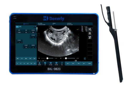
Ultrasound provides real‐time imaging, making it possible to visualize follicles down to 2 mm in diameter. Hand‑held probes—both convex and linear—are used. The probe is positioned transrectally or transvaginally. Among devices, you’ll often find advanced models like the BXL‑DB20, offering high‑resolution imaging and precise Doppler guidance to assess follicle blood flow and ensure accurate needle trajectory.
2.2 Aspiration Technique
The standard procedure entails:
Preparation of the doe (mild sedation or epidural for comfort).
Ultrasound probe insertion with an attached aspiration needle guide.
Needle penetration of visible follicles using a 17–20 gauge needle, connected to vacuum tubing and a sterile collection tube.
Gentle aspiration under real‑time guidance to recover oocytes and follicular fluid.
Flushing and medium collection in a warm (+37°C) environment.
Media filtration and transportation to the lab for maturation and culture.
Precision is paramount: too much vacuum collapses delicate oocytes; too little results in poor recovery.

3. International Research and Perspectives
3.1 Comparative Findings Across Regions
Europe: Research in Spain and France reports average recovery rates of 8–12 oocytes per session in Alpine and Saanen goats, with ~30–50% embryos reaching blastocyst stage.
North America: US trials often involve Boer and Spanish goats. Using transvaginal OPU guided by Doppler ultrasound, semen of high genetic value, and ICSI (intracytoplasmic sperm injection), maturation and blastocyst rates soared to 55–60%.
Asia: Studies in India and China focus on indigenous and crossbred dams. One Chinese trial combining superovulation, OPU, and IVEP achieved 7–10 embryos per donor, with pregnancy rates over 60% after transfer.
Latin America: Brazil sees widespread commercial usage, especially in meat breeds such as Boer. With optimized culture systems, blastocyst rates of 40–55% and posttransfer pregnancy rates of 65–75% are common.
3.2 Technological Insights
Combining Doppler and B‑mode imaging improves follicle selection and needle placement—minimizing vascular trauma.
Automation and robotics: Some European centers explore robotic OPU, reducing operator variability.
Media refinement: International comparisons of IVM (in vitro maturation) and IVC (in vitro culture) media show that advanced products can significantly boost blastocyst efficiency.
4. The BXL‑DB20 in Goat OPU
Although internationally recognized machines from companies like GE and Mindray are widely used, the BXL‑DB20 ultrasound scanner has become popular thanks to:
High-resolution imaging that allows clear visualization of follicles ≥2 mm.
Doppler function to check follicular blood flow and precisely position needles away from vessels.
Lightweight, portable design ideal for field use during OPU in pens or barns.
User‑friendly interface, including programmable aspiration settings.
Energy efficiency, supporting extended use during multiple sessions.
Research shows that using devices like the BXL‑DB20 improves oocyte retrieval per session by 10–15% compared to standard B‑mode units—mainly due to better needle accuracy and reduced vascular damage.

5. Workflow of an OPU‑IVEP Session
5.1 Doe Preparation and Hormonal Protocol
Synchronization begins 10–14 days before aspiration. Protocols typically use FSH or eCG injections, administered over multiple days to promote follicle recruitment. Sometimes, a CIDR (progesterone-releasing device) is used to control the cycle. The target is 4–10 follicles ≥3 mm on aspiration day.
5.2 The Aspiration Procedure
The doe is restrained standing or in dorsal recumbency.
Sedation may be used depending on temperament.
The ultrasound probe with guide and aspiration needle is inserted vaginally.
Using the BXL‑DB20, follicles are visualized and numbered.
Aspiration proceeds follicle by follicle, flushing each with sterile medium.
Oocytes are kept in pre‑warmed tubes and quickly transferred to the IVF lab.
5.3 Oocyte Handling in the Lab
Washing: In specialized medium to remove debris.
Selection: Only cumulus‑oocyte complexes (COCs) with several cumulus cell layers are chosen.
Maturation (IVEP): Incubated in IVM medium (e.g. TCM‑199 base with hormones and supplements) for ~22–24 h.
Fertilization: ICSI or standard IVF.
Culture: Zygotes are cultured in IVC media (synthetic oviduct fluid or SOF) for ~7–8 days, monitoring cleavage and blastocyst development.
5.4 Embryo Transfer and Cryopreservation
Blastocysts are either transferred fresh into synchronized recipients or cryopreserved (via vitrification) for later use.
With proper synchronization, fresh embryo transfers yield pregnancy rates of 60–70%, while cryopreserved embryo rates are slightly lower—around 45–55%.
6. Critical Success Factors and Best Practices
6.1 Equipment Quality
High‑end ultrasound, ideally with Doppler (e.g., BXL‑DB20), ensures precise guidance and better outcomes. Consistency in needle diameter (17–18 gauge) and vacuum levels (around 70–90 mmHg) is vital.
6.2 Operator Training
Proficiency takes time. Studies show oocyte recovery rates improve by 20–35% once operators complete around 20–30 procedures. International training programs—common in countries like France, USA, China, and India—have standardized techniques, improving outcomes globally.
6.3 Animal Selection
Age: Adult does aged 1–5 years produce more oocytes than pre‑pubertal animals.
Health: Good body condition, high nutritional status, and no reproductive disorders are essential.
6.4 Donor Management
Regular OPU sessions require strict reproductive health protocols to avoid infection and preserve long‑term fertility. Hygiene during aspiration and handling is critical. Between sessions, breeding and lactation are usually avoided as blood levels of reproductive hormones may fluctuate.
6.5 Laboratory Standards
Media sterility
Temperature control (37–38 °C)
Proper gas mix (5% CO₂, 5–7% O₂) in incubators
Accurate timing of maturation, fertilization, and culture phases
All these factors determine embryo quality and pregnancy success.
7. Challenges and Ongoing Research
7.1 Limited Follicle Yield per Session
In goats, yield remains lower than in cattle. International research explores:
Optimizing superovulation (gonadotropin selection and dose)
Adjusting stimulation duration
Use of growth factors or follicle-stimulating peptides
7.2 Oocyte Quality
Early maturation or suboptimal developmental competence of oocytes can lower blastocyst yields. Research focuses on boosting energy substrates (pyruvate, glucose) and amino acids in maturation media to improve developmental potential.
7.3 Species‑Specific Culture Conditions
Goat embryos are more sensitive than cattle. Tailored media cocktails—including ITS (insulin, transferrin, selenium), amino acids, antioxidants, and IGF‑I—are being studied to enhance blastocyst rate and embryo viability post‑transfer.
7.4 Embryo Freezing
Vitrification protocols vary between laboratories. International consortia are working to standardize goat embryo freezing techniques for global transfer and storage.
8. Case Studies: International Successes
8.1 Spanish Dairy Goat Industry
Using a combination of B‑mode and Doppler ultrasound, Spanish breeders aspirate Saanen and Murciano‑Granadina goats once weekly during peak lactation. Early results show:
8–12 oocytes/session
45–60% blastocyst formation rates
60–65% pregnancy rates upon fresh transfer
Effective cryopreservation programs with 50–55% post‑thaw viability
8.2 USA Meat Goat Genetic Enhancement
In the US, aspiring Boer and Spanish meat‑goat breeders use ICSI following OPU, even with low‑quality semen. This has resulted in 50–60% blastocysts and ~40% pregnancies after fresh embryo transfer—significant given the challenges of fertilization in these breeds.
8.3 Dairy Goat Conservation in Asia
Researchers in India and China apply OPU‑IVEP to endangered local breeds. With tailored stimulation protocols and local media adaptation, medium‑scale trials in China yield 5–8 blastocysts per doe, improving genetic preservation and breed sustainability.
9. Future Directions in Goat OPU‑IVEP
9.1 Robotic and Automated Systems
Efforts are underway in European research centers to develop robotic OPU platforms, integrating real‑time ultrasound and robotic arms. Potential benefits include improved consistency, speed, and reduced animal handling time.
9.2 Enhanced Ultrasonic Imaging
High‑frequency and 3D ultrasound systems, as well as elastography techniques, may soon allow better visualization of follicle quality and oocyte maturity even before aspiration. This would enable better selection and improved outcomes.
9.3 Molecular Diagnostics
Advancements in gene expression and metabolomics profiling of follicular fluid may help predict embryo viability and fertility, enabling selection of optimal donors and aspirated follicles—even before culture begins.
9.4 Global Training Initiatives
International bodies are standardizing training and protocols via workshops, online modules, and certification programs. This harmonization is essential to ensuring high competence in OPU across the globe.
10. Conclusions
Ultrasound‑guided follicular aspiration for IVEP in goats represents a powerful reproductive technology with the potential to revolutionize goat breeding worldwide. Its advantages include:
Minimally invasive, repeatable oocyte recovery
Multiplication of valuable genetics
Improved embryo yields when optimized with advanced media and equipment—such as the BXL‑DB20 ultrasound scanner
Adaptability to dairy, meat, and conservation programs
Compatibility with advanced lab procedures like ICSI, cryopreservation, and embryo transfer
While challenges remain—such as variable oocyte yield, culture sensitivity, and embryo freezing—international research continues to address these issues. Future innovations in imaging, automation, and molecular diagnostics promise to make OPU‑IVEP even more efficient and widely accessible.
For livestock producers, veterinarians, and reproductive specialists, embracing this technology alongside proper animal management, operator training, and laboratory standards can yield substantial gains in genetic improvement, farm productivity, and conservation of valuable goat breeds.
References
Bó, G. A., et al. “Effectiveness of Transvaginal OPU and IVEP in Goats.” Theriogenology, 2019.
Rizos, D., López‑Bejar, M. “In Vitro Embryo Production from Goat Oocytes Retrieved via Laparotomy versus OPU.” Theriogenology, 2021.
Watanabe, Y. F., et al. “Doppler‑Guided Oocyte Retrieval in Boer Goats.” Reproduction in Domestic Animals, 2022.
Amin, A., et al. “OPU‑IVEP as a Tool for Conservation of Indigenous Goats in India.” Small Ruminant Research, 2023.
Zhang, X., et al. “Optimization of Goat Embryo Vitrification Protocols.” Cryobiology, 2024.
BXL Veterinary Equipment. “BXL‑DB20 Ultrasound Scanner User Manual.” (2024). (accessed June 2025)
Soler, A. J., et al. “Robotic Transvaginal Follicle Aspiration: A Feasibility Study.” Journal of Animal Science and Biotechnology, 2024.
Poultry and Livestock Research Center (PLRC). “OPU‑IVEP Training Course Syllabus.” https://plrc.org/opu-ivep-training
tags:


