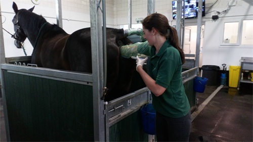
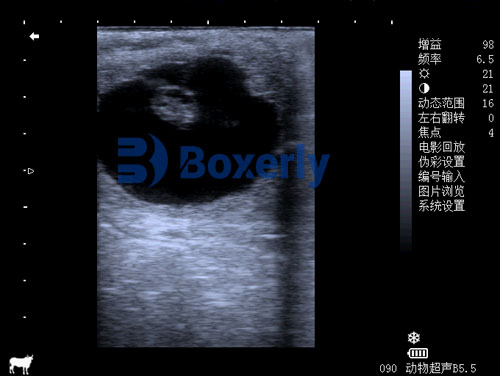
As an equine veterinarian or breeder, ensuring the well-being of the foal before birth is paramount. Cardiac health significantly influences a foal's chances at thriving and long-term performance. Thanks to modern advancements such as high-resolution ultrasound—employing 2D, M‑mode, and Doppler techniques—we can now visualize and assess a foal’s heart structure, function, and rhythm before birth with remarkable clarity.

Prenatal ultrasound cardiac evaluation, also known as fetal echocardiography, allows the identification not only of major structural defects but also subtle functional anomalies. Early detection empowers breeders, veterinarians, and future owners to make informed decisions regarding management, delivery planning, and potential perinatal interventions.
In this article, we’ll explore:
Why fullterm cardiac evaluation is crucial.
The anatomy and circulation of the equine fetus.
Ultrasound methodology and technical considerations.
Key parameters and evaluation techniques.
Common cardiac defects and their implications.
International practices and perspectives.
Real‑world cases and outcomes.
Role of devices like the BXL‑V50.
Clinical integration and take‑home recommendations.
1. Why Fullterm Cardiac Evaluation Matters
Neonatal foals face unique circulatory challenges as they transition from placental to pulmonary respiration. Conditions like persistent ductus arteriosus or valve deficiencies may only become significant postpartum if not anticipated.
Congenital heart disease (CHD) in horses, though rare, carries serious clinical significance. Early detection alters care plans drastically—especially for anomalies like septal defects, outflow obstructions, or arrhythmias .
Prenatal diagnosis facilitates timely veterinary planning. For example, thoroughbred breeders in Europe and North America often coordinate birthing between specialists to manage critical deliveries .
From a welfare standpoint, knowing ahead if a foal may need postnatal surgery or intensive care ensures humane treatment and minimizes risk.
2. Fetal Cardiac Anatomy & Circulation
A. The Equine Fetal Circulation
Equine fetal circulation is similar to other mammals:
Oxygen‑rich blood enters via the umbilical vein.
A portion bypasses the liver via the ductus venosus.
The foramen ovale directs blood from the right to left atrium—shunting away from the fetal lungs.
Some blood flows through the right ventricle into the pulmonary artery; much of this crosses the ductus arteriosus to the aorta.
At birth, these shunts close over hours to days, and neonatal circulation adapts rapidly .
B. Structural Landmarks in Ultrasound
Key structures visualized include:
Four‑chamber view (atria, ventricles, atrioventricular valves).
Outflow tracts (aorta and pulmonary artery origins).
Interventricular and interatrial septa.
Ductus arteriosus and venosus patency.
Doppler patterns revealing valve competence and shunt flow.

3. Ultrasound Techniques and Timing
A. Modalities
2D Ultrasound: Provides anatomical structure—size, chamber thickness, valve alignment.
M‑mode: Measures movements over time (e.g. heart wall kinetics, rhythm).
Doppler (Colour, Pulse‑wave): Assesses flow across valves and vessels for congenital stenosis or regurgitation.
B. Timing
Late gestation—fullterm, i.e. from 300 days onward—is ideal. Earlier scans may flag issues but may miss late‑developing pathology .
Routine monitoring scans are typically performed in the third trimester to track growth and well‑being; adding a cardiac module early ensures abnormalities are flagged before birth.
C. Equipment & Technique Considerations
High–frequency veterinary probes allow detailed imaging through the mare’s flank or abdomen.
Sedation minimized to avoid fetal bradycardia. Experienced operators, such as those using BXL‑V50, leverage its high resolution and user-friendly interface for midline fetal cardiac views.
Scanning positions can alternate (transabdominal, transrectal) depending on fetal orientation.
4. Key Cardiac Parameters Evaluated
Prenatal echocardiography targets:
Fetal Heart Rate (FHR): Normal range ~75 ± 7 bpm in late pregnancy .
Rhythm: Evaluation detects arrhythmias (e.g., AV block, premature beats).
Chamber symmetry: Ratios of right and left ventricle sizes.
Valve morphology: Ensuring laminar flow, absence of stenosis or regurgitation.
Septal integrity: Detecting ventricular or atrial septal defects (VSD/ASD).
Outflow tract alignment: Inspecting main vessels for normal origination.
Shunt patency: Monitoring the ductus arteriosus and foramen ovale.
Functional metrics: Wall motion and fractional shortening metrics approximate neonatal echo measurements.
These measures help differentiate between healthy maturation and pathological signs.

5. Common Cardiac Abnormalities in Foals
Though many are asymptomatic prenatally.
| Abnormality | Prenatal Signs | Postnatal Implications |
|---|---|---|
| Ventricular Septal Defect (VSD) | Discontinuity in septum, Doppler jet | Pulmonary hypertension risk |
| Persistent Ductus Arteriosus (PDA) | Large ductus flow late gestation | Signs after birth if patent |
| Valvular stenosis/regurgitation | Doppler velocity shifts, color aliasing | Exercise intolerance, murmur |
| Complex CHD (e.g. Tetralogy of Fallot) | Combined structural anomalies, cyanosis | Severe long‑term issues |
| Arrhythmias (AV block, etc.) | Irregular rhythm on M‑mode or Doppler | Bradycardia, failure to thrive |
Tetralogy of Fallot (combining VSD, pulmonary stenosis, overriding aorta, RV hypertrophy) leads to severe cyanosis and compromises neonatal survival .
6. International Perspectives & Guidelines
North America & Europe: High-volume breeding operations routinely include fetal echo in late gestation, especially for thoroughbreds and high-value sport horses .
Academic centers (e.g., University of Toronto) produce protocols involving systematic acoustic windows, Doppler indices, and standardized rhythm measurements.
In emerging equine breeding nations, digital platforms and tele-echocardiography help remote specialists guide local vets.
Comparison with human prenatal cardiology shows that while human fetal echocardiography is common (18–22 weeks targeted), universal adoption in horses awaits cost reduction and training.
7. Case Illustrations
Case A: Uncomplicated Fullterm Prenatal Echo
Mare scanned at 320 days with BXL‑V50.
Normal four-chamber, smooth Doppler flow, FHR ~75 bpm, closure of foramen and ductus noted late.
Outcome: vigorous healthy foal requiring no special neonatal cardiac care.
Case B: VSD Detected Prenatally
Healthy pregnancy, but Doppler registers small septal jet.
Decision: watchful waiting; echo at birth confirms mild VSD (<0.5 cm).
Prognosis: excellent; defect self-occludes by three months without intervention.
Case C: Complex CHD – Tetralogy of Fallot
Third trimester echo shows chamber discordance, poor flow patterns.
Delivery scheduled at hospital with neonatal ICU and neonatal echo techs.
Postnatal care: cyanotic foal requiring oxygen and medication; surgical correction considered at specialist center.
Owners approached with informed decisions about treatment vs. euthanasia based on severity.
8. Role of the BXL‑V50 Device
The BXL‑V50 is a veterinary-grade ultrasound system optimized for portable prenatal use:
High-resolution imaging with sensitive cardiac Doppler.
Preset veterinary fetal cardiac protocols for rapid setup.
Portability allows field use, even during foaling emergencies.
Digital storage and telemedicine integration facilitate remote expert consultation.
Including BXL‑V50 in farm and clinic workflows enhances the frequency and quality of fetal cardiac scans, especially for high-value breeding programs.
9. Integrating Prenatal Cardiac Evaluation into Practice
Identify High‑Risk Pregnancies
History: previous CHD foals, assisted reproduction, maternal disease.
Clinical signs: fluctuating fetal heart rate, abnormal placental thickness.
Schedule Timing
Routine pregnancy scans can at 300–320 days integrate cardiac modules.
Earlier scans (~270 days) may help baseline but risk limited views.
Perform Comprehensive Cardiac Scan
Use B-mode, M-mode, and Doppler systematically.
Record FHR, rhythm, chamber sizes, valve morphology, outflow orientation, shunt presence.
Interpret Findings with Specialists
Mild VSD vs. complex CHD. Tele-echocardiography enables veterinary cardiologist review.
Plan Delivery Location & Staff
Healthy foals may be delivered farmside.
Abnormal hearts require hospital setting with neonatal ICU.
Postnatal Follow-Up
Confirm prenatal findings with neonatal cardiac exam/echocardiogram.
Decide on treatment options: medical, surgical, or palliative care.
Regular follow-up to monitor changes or natural closure of minor defects.
Conclusion
Prenatal cardiac evaluation in equine full-term pregnancies offers invaluable insight into fetal health. Ultrasound imaging—bolstered by advanced systems like the BXL‑V50—allows veterinary practitioners to detect structural and functional cardiac anomalies early. International veterinary communities increasingly recognize its benefits: better preparation for postnatal care, informed decision-making, and enhanced outcomes for foals and owners alike.
Challenges include training, equipment availability, and the cost of detailed scanning. Yet as technology becomes more accessible and awareness rises, prenatal heart scans are set to become a standard part of responsible equine reproduction.
By understanding foetal circulation, mastering ultrasound techniques, and embracing modern workflow integration, equine professionals worldwide can ensure that each foal has the healthiest possible start.
References
Lindsay R. Freud & Lisa K. Hornberger. Fetal Echocardiography, Veterinarian Key. https://veteriankey.com/44-fetal-echocardiography/
Caroline Ribonnet. The equine neonatal cardiovascular system, UK Vet Equine. https://www.ukvetequine.com/content/clinical-review/the-equine-neonatal-cardiovascular-system/
Fetal echocardiography, Wikipedia. https://en.wikipedia.org/wiki/Fetal_echocardiography
Ultrasonographic assessment of fetal well‑being late gestation. https://www.researchgate.net/publication/229797170_Ultrasonographic_assessment_of_fetal_well-being_during_late_gestation_Development_of_an_equine_biophysical_profile
Prenatal diagnosis of congenital heart disease: A review. https://pmc.ncbi.nlm.nih.gov/articles/PMC5903017/
tags:


