For horse breeders and equine veterinarians, the final stages of a mare's pregnancy are both exciting and nerve-wracking. The anticipation of a healthy foal is always accompanied by a sense of uncertainty. Will the foal be fully developed? Is its growth on track? Are there any hidden complications? In recent years, equine ultrasonography—especially during late gestation—has become a highly valuable, non-invasive tool to assess fetal development and support decision-making before parturition. In this article, I’ll share how ultrasound is used to monitor fullterm foal development, what parameters we assess, and why this technique is increasingly essential for equine breeding programs worldwide.

Understanding Late Gestation in Mares
The gestation period of a horse averages around 340 days, though some variation exists between breeds and individuals. The last trimester, especially the final month, is critical for fetal development. During this phase, the majority of fetal weight gain occurs, with organs maturing and the musculoskeletal system strengthening in preparation for birth.
In the past, veterinarians relied largely on external signs, palpation, and the mare's history to estimate fetal well-being. However, these methods provide limited information about the fetus itself. With the growing availability of portable, high-resolution ultrasound devices, we can now visualize the foal inside the uterus and measure specific anatomical features that offer critical insights into its growth status.
Why Ultrasound? The Modern Approach
Ultrasound, particularly B-mode ultrasonography, has revolutionized equine prenatal care. Unlike X-rays or invasive procedures, ultrasound is safe, painless, and repeatable throughout gestation. This makes it ideal for monitoring fetal development, detecting abnormalities, and even predicting delivery complications.
Veterinarians in countries like the USA, Australia, the UK, and several European nations have incorporated routine late-gestation ultrasound scans into standard breeding management. The benefits are substantial:
Non-invasive and safe for both mare and fetus
Real-time assessment of fetal development
Identification of high-risk pregnancies before clinical signs appear
Accurate measurements to evaluate fetal maturity
For commercial breeders, this technology is not just about curiosity; it’s a practical tool that helps optimize foal survival rates, minimize losses, and improve long-term herd productivity.
Key Ultrasonographic Parameters Measured in Late Gestation Foals
During the final stages of gestation, several fetal measurements are commonly obtained via transabdominal or transrectal ultrasound:
1. Fetal Eye Orbit Diameter
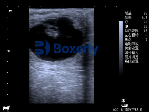
The measurement of the fetal eye orbit is one of the most validated parameters in late gestation. Numerous studies, such as those published by Woods et al. (2020) in the Journal of Equine Veterinary Science, have established normative growth charts correlating eye orbit size with gestational age and fetal health. An unusually small or large orbit may indicate intrauterine growth restriction (IUGR) or developmental anomalies.
2. Biparietal Diameter (BPD)
BPD refers to the distance between the parietal bones of the fetal skull. This measurement offers another reliable index of fetal age and development. In many equine hospitals, BPD is regularly monitored starting from mid-gestation, but it remains informative even in the final weeks.
3. Fetal Heart Rate
By evaluating the fetal heart rate, we can gain insight into the foal's current well-being. A persistently elevated or depressed heart rate may signal fetal distress, hypoxia, or placental insufficiency, prompting closer monitoring or early intervention.
4. Placental Thickness and Appearance
In addition to evaluating the fetus itself, ultrasound allows for thorough assessment of the placenta. Thickened placenta or evidence of placental separation can be early indicators of conditions like placentitis—a common cause of late-term pregnancy loss in mares.
5. Fetal Movement
Active movement patterns, observed via real-time imaging, are also used as indirect indicators of fetal well-being. Decreased activity may suggest compromised oxygen delivery or infection, while excessive movements can sometimes precede dystocia.
The S-Shaped Growth Curve in Foal Development
Like many mammals, foals exhibit an S-shaped growth curve during development. In early gestation, cellular differentiation dominates, followed by rapid organogenesis and skeletal formation. As the pregnancy progresses, growth accelerates sharply during the third trimester, plateauing as fullterm approaches.
Ultrasound measurements taken during late gestation allow breeders to verify whether the fetus is following this expected curve. In cases where growth plateaus too early or lags behind established benchmarks, targeted interventions such as modified nutrition, pharmacological support, or increased monitoring may be warranted.

Clinical Applications in Different Breeding Scenarios
Across various countries, equine breeders have integrated late gestation ultrasound into different breeding and management strategies:
1. High-Value Breeding Programs
In Thoroughbred racing, Standardbred performance horses, and warmblood sport horse breeding programs, every foal represents significant financial and genetic investment. In these high-stakes environments, breeders in the USA, UK, and Germany regularly schedule ultrasound examinations during weeks 300–320 of gestation to confirm fetal readiness for birth and prepare for potential complications.
2. Managing High-Risk Mares
Certain mares carry higher risks of pregnancy complications, such as those with a history of placentitis, prior stillbirths, twin reduction procedures, or metabolic disorders like equine metabolic syndrome. For these individuals, veterinarians in Australia and Canada often recommend weekly or bi-weekly late gestation ultrasound scans to track fetal status and placental integrity closely.
3. Predicting Premature or Prolonged Labor
In some cases, ultrasound can help identify signs that suggest premature delivery risk or prolonged gestation. For example, insufficient fetal size, abnormal fluid levels, or placental abnormalities may influence the veterinarian’s decision to intervene early or delay induction.
4. Supporting Foaling Preparation
By confirming fetal maturity through ultrasound indicators (normal eye orbit size, adequate skeletal ossification, active movement, stable heart rate), breeders can better plan for foaling assistance, neonatal care, and emergency preparedness, significantly improving foal survival outcomes.
Technological Advances: The Role of Portable Ultrasound
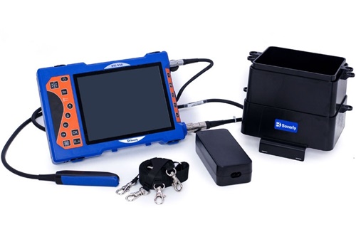
The evolution of portable, rugged ultrasound systems has made late gestation assessment more accessible, even in remote or field settings. Devices like the BXL-V50 Veterinary Ultrasound Scanner, with high-definition displays, waterproof design, and multi-probe capability, allow for detailed fetal imaging directly on breeding farms.

Many veterinarians favor such portable systems because:
They minimize stress by scanning mares in familiar surroundings.
They provide high-quality imaging comparable to stationary hospital-grade units.
They support both transabdominal and transrectal scanning depending on the stage and patient condition.
They enable real-time data sharing with referral specialists through digital storage and telemedicine features.
In the USA, Europe, and parts of Asia, this portability has dramatically increased the adoption of routine late gestation scanning, even among small and mid-sized breeders.
Research Supporting Late Gestation Ultrasound Use
The scientific literature strongly supports the value of late gestation ultrasound in horses. For example:
Woods, G. L., et al. (2020) demonstrated how consistent monitoring of fetal eye orbit diameter improves prediction of gestational age and reduces perinatal mortality.
Renaudin, C. D., et al. (2019) examined placental thickness measurement as an early indicator of placentitis, allowing proactive treatment.
Kutzler, M. A. (2018) outlined comprehensive guidelines for ultrasonographic fetal evaluation in equine practice, now widely adopted in North American veterinary teaching hospitals.
These studies confirm that ultrasound offers both predictive and diagnostic value during this critical phase of equine reproduction.
Limitations and Considerations
While ultrasonography offers tremendous benefits, it is not without limitations:
Operator Skill Dependency: Accurate measurements require experience and technical expertise.
Fetal Positioning: Certain fetal positions may obscure key structures.
Equipment Limitations: Lower-resolution or outdated machines may not provide the image quality necessary for fine assessments.
Thus, ongoing training, proper equipment maintenance, and awareness of scan limitations remain crucial for maximizing the diagnostic value of ultrasound.

Future Perspectives
Looking ahead, many equine reproductive specialists envision even greater reliance on advanced imaging technologies. Three-dimensional ultrasonography, AI-assisted growth analysis, and remote fetal monitoring are being actively researched. These innovations aim to offer even earlier detection of developmental issues, better foaling predictions, and personalized reproductive management for each mare.
In some countries, collaborative research between veterinary schools and breeders has already begun refining breed-specific growth charts using AI-driven big data analysis of fetal ultrasound images. Such efforts will further standardize fetal growth expectations and optimize intervention timing.
Conclusion
For equine breeders around the world, ensuring a healthy foal at birth is both a science and an art. Late gestation ultrasound provides an unparalleled window into the final stages of fetal development, enabling veterinarians and breeders to detect problems early, optimize foaling management, and improve survival rates.
By carefully monitoring parameters such as eye orbit diameter, biparietal diameter, fetal heart rate, placental thickness, and movement patterns, we gain valuable data to support decision-making during this critical period. With advances in portable, high-resolution ultrasound technology like the BXL-V50, this powerful diagnostic tool is becoming more accessible than ever before—transforming equine breeding practices across the globe.
As awareness and technology continue to advance, late gestation ultrasound will undoubtedly remain a cornerstone of responsible, high-quality equine reproduction management for years to come.
Reference Sources
Woods, G. L., et al. (2020). "Equine Fetal Eye Measurements by Ultrasound: Establishing Growth Norms." Journal of Equine Veterinary Science, 89, 103097. https://www.j-evs.com/article/S0737-0806(20)30001-9/fulltext
Renaudin, C. D., et al. (2019). "Ultrasonographic Evaluation of Equine Placentitis." Equine Veterinary Education, 31(3), 138-146. https://onlinelibrary.wiley.com/doi/10.1111/eve.12835
Kutzler, M. A. (2018). "Ultrasonographic Monitoring of the Equine Pregnancy." Veterinary Clinics of North America: Equine Practice, 34(1), 67-85. https://www.sciencedirect.com/science/article/abs/pii/S0749073917301032
tags:


