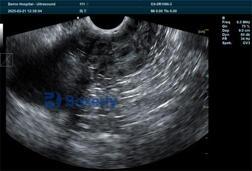
For small ruminant farmers and veterinarians, early and accurate pregnancy diagnosis plays a central role in optimizing reproductive efficiency, improving flock management, and maximizing profitability. Among the most reliable technologies available today, ultrasound scanning offers a non-invasive, real-time, and cost-effective solution to monitor reproductive status in animals such as sheep and goats. However, while ultrasound pregnancy detection has become increasingly popular worldwide, training veterinary professionals to competently operate ultrasound tools on small ruminants presents a unique set of challenges. In this article, we explore these challenges based on both practical experience and scientific literature, and discuss why mastering ultrasound skills remains crucial in modern small ruminant veterinary practice.

Understanding Ultrasound Use in Small Ruminants
Small ruminants, such as sheep and goats, are widely raised across the globe for meat, milk, wool, and fiber production. Because their reproductive efficiency directly influences farm economics, early pregnancy diagnosis is particularly valuable. Ultrasound, specifically real-time B-mode ultrasonography, allows for accurate pregnancy detection as early as 25–30 days post-breeding in sheep and goats.
In contrast to large livestock like cattle, small ruminants present specific anatomical and behavioral differences that complicate the training process. Their smaller body size, higher reproductive rates, and more delicate reproductive anatomy require veterinarians to develop advanced hand-eye coordination, image interpretation skills, and a nuanced understanding of reproductive physiology.
The Learning Curve of Ultrasound Training
Learning to use ultrasound equipment effectively on small ruminants involves mastering multiple technical and cognitive skills simultaneously:
Probe Handling and Positioning: Small ruminants have smaller pelvic cavities, making proper transabdominal or transrectal probe placement more challenging. Maintaining adequate contact with the animal's abdomen or rectum requires gentle yet firm handling to avoid causing discomfort or movement during scanning.
Image Acquisition: Unlike static imaging modalities, real-time ultrasonography requires practitioners to actively maneuver the probe to capture clear and interpretable images of the uterus, amniotic sacs, embryos, and placentomes. Novices often struggle with probe angulation and pressure adjustments, resulting in blurry or non-diagnostic images.
Image Interpretation: Identifying pregnancy structures (such as fluid-filled uterine horns, embryos, or fetal heartbeats) demands extensive anatomical knowledge and experience. Early gestational sacs may resemble other fluid accumulations, making misdiagnosis a risk for inexperienced operators.
Animal Handling and Stress Management: Small ruminants are prone to stress, which can affect both animal welfare and diagnostic accuracy. Handling animals calmly and safely during scanning is a skill that must be incorporated into ultrasound training programs.
As highlighted by international veterinary training institutions, achieving competency typically requires a combination of theoretical education, supervised practical sessions, repeated scanning practice on live animals, and continuous feedback from experienced instructors.
Species-Specific Training Difficulties
The anatomical differences between sheep and goats introduce additional training challenges:
Sheep: Fat-tailed or heavily fleeced breeds may have significant wool coverage that interferes with probe contact. Trimming wool, applying sufficient coupling gel, and positioning the animal correctly are crucial steps that must be learned.
Goats: Compared to sheep, goats often have more active gastrointestinal movement and deeper abdominal fat layers, which can obscure early pregnancy signs on ultrasound. Beginners frequently struggle with distinguishing digestive content from uterine fluid during the early stages of gestation.
International studies suggest that veterinary students and farm technicians learning ultrasound pregnancy diagnosis on small ruminants often require significantly more supervised practice sessions than when learning on larger animals like cattle.

Training Methods: Simulation vs. Live Practice
Several training methods have been developed globally to address the steep learning curve in small ruminant ultrasound training:
Didactic Instruction: Traditional classroom sessions cover the basics of reproductive anatomy, ultrasound physics, image interpretation, and common pathologies.
Simulation Models: Some veterinary schools have adopted ultrasound simulation phantoms, which allow students to practice probe handling and image acquisition without risking harm to live animals. However, simulators often fail to replicate the variability and unpredictability of live animal scanning.
Live Animal Practice: Direct supervised practice on live sheep and goats remains the gold standard for mastering scanning skills. However, ethical concerns, animal welfare regulations, and logistical constraints limit the availability of sufficient training animals in many countries.
Online Training Modules: With advances in digital education, online ultrasound courses with annotated videos and remote mentoring programs have become increasingly popular, especially in regions where experienced trainers are scarce.
Studies from North America, Europe, and Australia consistently emphasize that hands-on live animal experience, combined with expert mentorship, is the most effective way to overcome initial training difficulties and build confidence in ultrasound-based pregnancy diagnosis.
Common Errors During Early Training
In practice, novice ultrasound operators often encounter several predictable errors when scanning small ruminants:
False Positives: Misidentifying fluid-filled intestines or bladder structures as pregnancy sacs.
False Negatives: Failing to detect early pregnancies due to suboptimal probe positioning or lack of operator confidence.
Gestational Age Miscalculations: Incorrectly estimating fetal age due to poor image quality or misunderstanding fetal measurements.
Operator Fatigue: Scanning multiple animals in a short period often leads to deteriorating image quality due to physical fatigue and declining focus, a problem commonly reported in commercial flock scanning operations.
Addressing these issues requires both technical coaching and reinforcement of diagnostic protocols, such as scanning at standardized days post-breeding and confirming doubtful diagnoses during re-scans.
The Role of Modern Ultrasound Equipment in Training
Advances in Veterinary ultrasound technology have significantly eased some training challenges:
Portability and Durability: Modern portable devices allow trainers and students to scan animals directly in the field, where they are most comfortable.
High-Resolution Imaging: Devices with superior resolution provide clearer images, helping beginners identify pregnancy structures more easily.
Preset Modes for Small Ruminants: Some machines offer species-specific presets that automatically optimize image parameters for sheep and goats, reducing setup errors.
Data Storage: The ability to record and review scans later for teaching and feedback purposes improves learning outcomes.
For example, veterinary ultrasound systems such as the BXL-V50 have gained popularity worldwide for their robust design, extended battery life, and high-definition imaging that greatly benefit both novice and experienced operators during pregnancy diagnosis training.

Global Perspectives on Training Approaches
Training approaches to ultrasound pregnancy detection on small ruminants vary significantly across different regions:
Europe: In countries like the UK and France, structured certification programs for professional sheep scanning technicians include rigorous training and assessment. Some nations require licensure to ensure scanning competency and prevent misdiagnosis that could financially harm producers.
North America: Many veterinarians receive ultrasound training as part of postgraduate continuing education programs. Private companies also offer specialized small ruminant ultrasound workshops for both veterinarians and experienced farm staff.
Australia and New Zealand: Large-scale commercial sheep scanning operations have driven the development of highly specialized ultrasound scanning technicians who often scan thousands of ewes per season. Training programs emphasize speed and efficiency alongside diagnostic accuracy.
Developing Regions: In parts of Africa and Asia, limited access to trained instructors and affordable equipment remains a major barrier to widespread adoption of ultrasound pregnancy diagnosis. International veterinary organizations have initiated training outreach programs to build local capacity.
These global experiences highlight the universal need for comprehensive, practical, and accessible training systems to meet the growing demand for small ruminant ultrasound diagnostics.
Economic Importance of Ultrasound Training
For both farmers and veterinarians, the economic benefits of accurate ultrasound pregnancy diagnosis are clear:
Early Identification of Non-Pregnant Animals: Allows for timely rebreeding, sale, or culling decisions, reducing feed and labor costs.
Better Nutritional Management: Adjusting feeding programs based on pregnancy status and fetal numbers improves maternal health and lamb survival.
Optimized Labor Planning: Knowing expected lambing dates enables better scheduling of staff during peak birthing periods.
Enhanced Flock Genetics: Selective breeding based on reproductive performance data supports long-term genetic improvement programs.
However, these benefits are only fully realized when ultrasound operators are properly trained to deliver reliable diagnostic results. Poorly trained personnel risk making costly mistakes that undermine the value of the technology.

Recommendations for Improving Training Outcomes
Based on international research and field experiences, several key recommendations emerge for improving ultrasound pregnancy training on small ruminants:
Start with clear theoretical instruction before hands-on practice.
Use simulation models to build initial probe handling confidence.
Provide extended supervised practice sessions on live animals.
Emphasize the importance of animal handling and welfare during scanning.
Incorporate video-based case reviews and remote mentoring programs.
Offer regular continuing education opportunities to maintain and refine skills.
Encourage certification programs to establish recognized competency standards.
Conclusion
Ultrasound pregnancy diagnosis offers transformative benefits for small ruminant production worldwide, but the road to developing competent veterinary ultrasound operators is challenging. The unique anatomical and behavioral characteristics of sheep and goats require targeted, hands-on training strategies that blend theory with practice.
Despite these challenges, continued investment in effective training programs, access to modern ultrasound equipment, and global knowledge exchange are gradually raising the standard of ultrasound diagnostics in small ruminant veterinary medicine. As more veterinarians, technicians, and farmers gain proficiency in ultrasound pregnancy detection, the full potential of this technology can be realized—improving animal welfare, enhancing reproductive performance, and boosting profitability across diverse production systems.
Reference Sources:
G.A. Silva Del Rio, et al. (2022). "Practical Applications of Ultrasonography for Reproductive Management in Small Ruminants." Journal of Veterinary Science and Technology. https://www.veterinaryscijournal.com/articles/practical-applications-ultrasonography-small-ruminants
P. González-Bulnes & A. Santiago-Moreno (2023). "Advances in Ultrasonographic Diagnosis of Pregnancy in Sheep and Goats." Theriogenology. https://www.sciencedirect.com/science/article/abs/pii/S0093691X23000052
Sheep Ultrasound Technicians Association (SUTA), UK (2024). "Professional Training Standards for Small Ruminant Pregnancy Scanning." https://www.sheepscanning.org.uk/training
tags:


