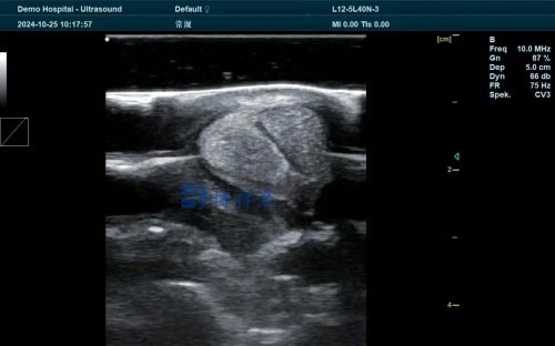
As global demand for animal protein rises, livestock reproduction must be both efficient and sustainable. Early, accurate pregnancy diagnosis is essential to maximizing reproductive output and minimizing costs. Veterinary ultrasound—particularly transrectal B‑mode and Doppler—has become widely adopted worldwide as a safe, non‑invasive, and reliable tool for early pregnancy detection, fetal monitoring, and managing reproductive health in cattle, sheep, goats, pigs, and other species.

In this article, we’ll explore how ultrasound is used in veterinary reproduction, focusing on its benefits, interpretation of scans, practical applications, and its global role in improving reproductive efficiency. I will also include foreign farmers’ and veterinarians’ perspectives on what makes ultrasound indispensable in modern animal husbandry.
1. Why ultrasound?
1.1 Global shift from palpation
Traditionally, pregnancy diagnosis relied on manual palpation—a method still used but with limitations. Ultrasound enables earlier detection—often at 25–30 days post‑breeding—where manual palpation reaches only about 45–60 days, and risks discomfort or injury.
1.2 Safety, precision, and repeatability
Ultrasound is non‑invasive, safe, portable, and quantitative. B‑mode ultrasound highlights fluid content and structures, while Doppler evaluates blood flow—helpful to assess corpus luteum function, early pregnancy viability, and estrous cycle status .
2. Core reproductive applications
2.1 Early pregnancy diagnosis
B‑mode imaging: At 20–28 days post‑insemination, the fluid‑filled uterus, heartbeat, and embryo sac can be detected reliably .
Doppler imaging: By assessing luteal blood flow at around day 20, practitioners can differentiate pregnant from non‑pregnant animals—achieving ~86–93% accuracy.
2.2 Fetal aging and sexing
Ultrasound enables fetal aging by observing key developmental milestones—heartbeat (~22 days), spinal cord (~28 days), limbs (~44 days), ribs (~52 days).
Fetal sexing is accurate (92–100%) between days 55–80 by identifying genital tubercle position.
2.3 Detecting twins and abnormalities
Transrectal scans at 40–55 days post-AI clearly show twin embryos, enabling selective management. Ultrasound also helps identify uterine or ovarian pathologies—such as cysts, abscesses, or prolapse—which manual palpation often misses.

3. Efficiency gains in management
3.1 Minimizing non‑productive intervals
Early diagnosis allows timely re‑synchronization and rebreeding of non‑pregnant animals, reducing calving intervals and increasing calf output per year.
3.2 Reducing feed costs
Identifying open cows early avoids unnecessary feed during winter—one American veterinarian estimates a saving of US $200–300 per non‑pregnant cow.
3.3 Improving herd structure
By diagnosing fetal sex and twin status early, producers can selectively breed, manage herd replacement, or prepare bi‑maternal facilities .
3.4 Enhancing herd health
Ultrasound aids in diagnosing reproductive disorders—ovarian cysts, endometritis, luteal dysfunction—enabling timely interventions and improved fertility .
4. Technical perspective
4.1 Equipment & probes
Portable veterinary units (5–9 MHz transducers) provide adequate resolution for uterine Structures; 5 MHz offers depth, 7.5 MHz greater clarity.
4.2 Operator skill & consistency
Accurate early detection depends on experience. Studies indicate accuracy reaches ~100% by day 20 when performed by experts. After initial training, ultrasound enhances overall palpation technique.
4.3 Use of Doppler
Color‑Doppler adds sensitivity by visualizing blood flow in corpus luteum or uterine arteries—particularly effective at day 20 for early pregnancy or luteal function assessment . It also helps identify appropriate embryo transfer recipients.
5. International applications & farmer experiences
5.1 United States
A USDA 2020 survey found only 32% of U.S. producers use pregnancy diagnostics. Dr. Folsom (Idaho) emphasizes cost savings from early non‑pregnant detection and fetal staging to streamline calving.
5.2 Australia & New Zealand
Australia’s Agriculture Victoria recommends ultrasound at 8–10 weeks post‑mating to reduce operator fatigue and accurately age fetuses for herd culling decisions.
5.3 Europe & Latin America
Spanish and Mexican studies highlight ultrasound’s diagnostic value for ovarian and uterine pathologies, as well as estrous cycle monitoring—extending herd health beyond reproduction.
5.4 Academic research
Institutional use in the U.S. and Europe drove early developments—University of Wisconsin pioneered follicular wave tracking in the 1980s, while current research continues to optimize Doppler protocols.

6. Best practices for implementation
6.1 Strategic timing
Initial scan: day 25–30 post‑AI. Follow‑up: day 60–70 to detect embryonic loss (15–16% loss between days 28 and 56) .
Tailor protocols to herd and species.
6.2 Training & equipment investment
Line up skilled technicians and invest in rugged, portable units. Doppler adds value for embryo transfer or cycle management.
6.3 Data‑driven decisions
Record outcomes—pregnancy rates, early losses, twin frequency, calving intervals. Match scanning with resynchronization or nutritional adjustments.
6.4 Counseling herd owners
Explain economic ROI—feed savings, improved conception rates, healthier calves. Share case studies: ID ranch saved ~$250 per dry cow; AU station improved calves weaned by 3%.
7. Limitations & challenges
7.1 Access and cost
Equipment and operator training pose barriers, especially for small farms. Mobile vet services may be needed.
7.2 Early pregnancy ambiguity
Before day 20, false negatives may occur; color Doppler helps but is not infallible.
7.3 Operator variability
Early detection depends heavily on skill. Ongoing training and standardization are crucial.
8. Future outlook
8.1 Technological innovations
AI‑driven image analysis may enable automated pregnancy scans with smartphones or drones—bringing ultrasound to remote areas.
8.2 Affordable Doppler
Portable color‑Doppler units are evolving; widespread adoption could make early pregnancy screening and cycle management more precise.
8.3 Global adoption
As awareness grows, more developing‑country producers could access ultrasound, boosting rural livelihoods and food security.

Conclusion
Veterinary ultrasound pregnancy diagnosis has ushered in a new era of reproductive management. Offering early detection, precise monitoring, and a host of indirect benefits—feed savings, better herd structure, and animal health—it has become globally essential.
Data-driven usage of ultrasound enables producers to make informed decisions: which animals to re‑breed, cull, or manage differently. Across geographies, expert farmers and vets alike attest to its power in optimizing production and welfare.
Looking ahead, ultrasound’s future lies in AI guidance, lower‑cost units, and global training programs—propelling livestock reproduction into a smarter, more sustainable future.
References
Fricke PM. Scanning the future – Ultrasonography as a reproductive management tool for dairy cattle. J Dairy Sci. 2002;85(8):1918–26. DOI:10.3168/jds.S0022-0302(02)74268-9
Poock SE, Wilson DJ. A review of the use of ultrasound for reproductive purposes in beef cattle. Proc Appl Repro Strat Beef Cattle. 2011.
IFAS, Univ. of Florida. Practical Uses for Ultrasound in Managing Beef Cattle Reproduction. 2 years ago.
Fontes PLP, Oosthuizen N. Applied use of Doppler ultrasonography in bovine reproduction. Frontiers Anim Sci. 2022.
Agriculture Victoria. Pregnancy testing of beef cattle. 1.3 years ago.
Quintela LA et al. Use of ultrasound in the reproductive management of dairy cattle. Reprod Dom Anim. 2012.
BovineVetOnline. Ultrasound technology helps veterinarian stage pregnancy... 8 months ago.
IMV Technologies. Use of ultrasound scanning technique – bovine. 3.4 years ago.
tags:


