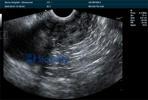
As a livestock producer, ensuring fetal viability and accurate gestational assessment is fundamental to herd management efficiency. Ultrasonography has emerged as a non-invasive, real-time, and reliable tool to monitor pregnancies in cattle, sheep, goats, and other production animals. In this comprehensive article, drawing on both international research and farm-level insights, we explore how ultrasound imaging predicts fetal viability in livestock, enhances reproductive decisions, and ultimately supports profitability and animal welfare.

1. Importance of Early and Accurate Pregnancy Diagnosis
Early detection of non-viable pregnancies minimizes unnecessary nutritional costs, allows prompt rebreeding, and reduces open days. Research shows embryonic mortality in cattle ranges from 6% to 25%, depending on timing and management. Using ultrasound to confirm fetal heartbeat and movement around day 22–30 post-insemination significantly improves differentiation between viable and non-viable pregnancies .
A comparative study by E.I. Medical Imaging found that using ultrasound instead of rectal palpation reduced fetal losses (1.29% vs. 2.68%) with experienced technicians. This aligns with findings from bovine practice, where ultrasound is considered safer and more reliable than palpation.
2. Key Ultrasound Techniques and Their Application
2.1 Transrectal Real‑Time B‑Mode Ultrasound
The standard field method involves transrectal, real-time B‑mode ultrasound. With a linear or convex probe, veterinarians detect:
Gestational sac and chorioallantoic fluid
Embryo presence and heartbeat
Crown–rump length to estimate gestational age
Fetal movement as a viability marker
Embryos are detectable as early as day 20–22, with a visible heartbeat. Chorioallantoic fluid landmarks assist in confirming early pregnancies.
2.2 Doppler Ultrasound
Color-flow Doppler ultrasound assesses luteal blood flow around day 20 post-AI. A well-vascularized corpus luteum independently predicts pregnancy with >90% accuracy. Doppler aids in:
Earlier diagnosis than B‑mode
Resynchronization protocols on day 20
Reducing unnecessary CIDR use and open periods
3. Predicting Fetal Viability: Indicators and Timetable
3.1 Fetal Heartbeat and Movement
Heartbeat confirmation (~day 22) and observable fetal movement (~day 45) are classic viability markers. In non-viable cases, absence of heartbeat, movement, or presence of echogenic debris like flocculent fluid (>day 75) suggests fetus degeneration.
3.2 Crown–Rump Measurements and Protocol
Anatomical dimensions measured by ultrasound allow precise gestational aging when conducted at specific intervals. For example, at day 35, crown–rump ~1 cm; day 50, ~4 cm—these metrics guide reproductive timelines . This precision is vital for protocols like super-early resynchronization.
3.3 Multiple Pregnancy Detection
Twinning in cattle brings management challenges. Ultrasound effectively identifies twin gestations, enabling targeted nutritional and calving plans. This early detection is key, as spontaneous reduction may occur before day 90.

4. Farm‑Level Practice and Equipment Considerations
4.1 Field-Friendly Devices
Veterinary ultrasound units designed for field conditions are portable, battery-powered, and robust. They often include:
Converted linear or convex transducers suited for transrectal use
Gridded overlays replacing calipers for basic measurement
Peak portability for outdoor application
4.2 Technician Skill and Protocols
Accuracy strongly correlates with operator experience. Novices show ~2.1% fetal loss versus ~1% by pros. Best practices include:
Scanning day 30–32 post-AI for reliable detection
Combining heartbeat, gestational sac, and luteal status
Maintaining clean but practical hygiene on field equipment
5. Economic and Management Advantages
Ultrasound’s practical benefits encompass:
Early rebreeding: Confirmed open cattle can be reinseminated swiftly, lowering calving intervals.
Resource optimization: Identifying failed pregnancies early avoids feeding non-pregnant animals.
Twin management: Enables tailored care, nutrition, and calving oversight.
Resynchronization programs: Doppler-assisted timing accelerates reproductive cycling.
Improved welfare: Non-invasive testing and quicker decisions reduce stress on cattle.
6. International Scenarios and Emerging Trends
6.1 Dairy Cattle
Studies in dairy herds show 20–45% pregnancy losses between fertilization and term. Early ultrasound (day 27–30) enables timely rebreeding and maintains sustainably high productivity.
6.2 Small Ruminants and Other Livestock
Ultrasound is also crucial in sheep, goats, and buffaloes. Its versatility extends to:
Pregnancy diagnosis
Fetal counting
Sexing in research contexts
6.3 Companion and Emerging Species
In dogs (bitches), ultrasound assists prediction of parturition date and litter size without harmful radiation. In camels and exotic livestock, field-adapted protocols are being developed .
6.4 Biomarkers and Proteomics
Research is progressing into pregnancy-associated glycoproteins (PAGs), early pregnancy proteins, and microRNA biomarkers in cows. These may further enhance early, non-imaging-based diagnostic accuracy.

7. Limitations and Risk Management
Technician skill: Inexperienced operators may misinterpret images or cause fetal loss.
Timing sensitivity: Scans before day 25 can misdiagnose early embryonic loss as pregnancy failure.
Equipment trade-offs: Field ultrasound sacrifices image resolution for portability .
False positives: Uterine fluid or pathologies may mimic pregnancy early on.
8. Best‑Practice Guidelines for Predicting Fetal Viability
| Stage | Time (Days Post‑AI) | Target Findings | Action |
|---|---|---|---|
| Early | 20–22 | Corpus luteum blood flow (Doppler) & fluid detection | Resynchronization or confirm setup |
| Mid | 28–32 | Gestational sac, embryo, heartbeat | Pregnancy or start rebreeding |
| Later | 45–60+ | Fetal movement, size for gestational monitoring | Twin detection, adjust nutrition |
| Outcome | All | Repeat scans to confirm viability | Prompt rebreeding if needed |
Conclusion
Veterinary ultrasound pregnancy imaging stands as a cornerstone of modern livestock reproduction management. By enabling early detection of pregnancy, verification of fetal viability, and monitoring of gestational progression, ultrasound empowers farmers and veterinarians to make data-driven decisions, reduce economic loss, and prioritize animal welfare. As technology evolves—integrating Doppler, biomarker testing, and portable imaging—the potential for precise, efficient breeding programs grows even stronger.
Embracing ultrasonography on your farm ensures not only productive operations, but also healthier animals and improved bottom lines. With webinars, veterinary training, and continuing research, global adoption is increasing. If you haven’t yet integrated this powerful tool, now is the time.

References
Beal W.E., Perry R.C., Corah L.R. The use of ultrasound in monitoring reproductive physiology of beef cattle. J Anim Sci. 1992;70(3):924–929. PMID 1564011.
Fricke P.M., Lamb G.C. Practical application of ultrasound in dairy cattle reproduction. J Dairy Sci. 2002;85(8):1918–26.
IMV Imaging. Confirmation of fetal viability in cattle. IMV Imaging Learning Resources. 2025.
E.I. Medical Imaging. Fetal Loss From Ultrasound vs. Palpation in Beef. Blog. Jan 2011.
IFAS, University of Florida. Practical Uses for Ultrasound in Managing Beef Cattle Reproduction. 2022.
Szenci O. Recent Possibilities for the Diagnosis of Early Pregnancy and Embryonic Mortality in Dairy Cows. Animals. 2021;11(6):1666.
Schrank M., Pereira M., Mollo A. Diagnostic imaging in the pregnant bitch: risks, advantages, and disadvantages. Front Vet Sci. 2025;12:1588847.
tags:


