In modern veterinary reproductive management, understanding the ovarian follicular reserve and its depletion over time is critical to optimizing breeding outcomes in aging livestock. Follicular reserve—the stockpile of primordial follicles within the ovary—dictates the reproductive lifespan and fertility potential of breeding animals. With advances in veterinary ultrasound technology, specifically high-resolution B-mode ultrasonography, veterinarians and animal scientists can non-invasively quantify follicular reserve depletion, enabling informed decisions on breeding strategies, animal replacement, and fertility preservation.

This article explores how veterinary ultrasound is used to evaluate follicular reserve depletion in aging breeding animals. Drawing from international research and practical farm experience, we examine the physiological basis of follicular dynamics, the technical ultrasound approaches for follicular quantification, and the implications for breeding program management.
Understanding Follicular Reserve and Its Importance in Breeding Animals
The ovarian follicular reserve is composed of primordial follicles formed before birth, which gradually decrease as animals age due to continuous recruitment and atresia. This reserve serves as the finite source for developing follicles capable of ovulation and fertilization throughout an animal’s reproductive life.
In livestock such as cattle, sheep, pigs, and goats, the depletion rate of follicular reserve varies by species, breed, nutrition, and environmental factors. As the follicular pool declines, reproductive efficiency diminishes, manifested by lower conception rates, prolonged intervals between estrus cycles, and increased early embryonic losses.
Traditionally, quantifying follicular reserve was challenging, relying on invasive histological techniques or indirect hormonal assays. However, veterinary ultrasound technology now allows real-time visualization of ovarian follicles, including small antral follicles that correlate closely with the underlying reserve.
Ultrasound Techniques for Follicular Reserve Quantification
B-Mode Ultrasonography
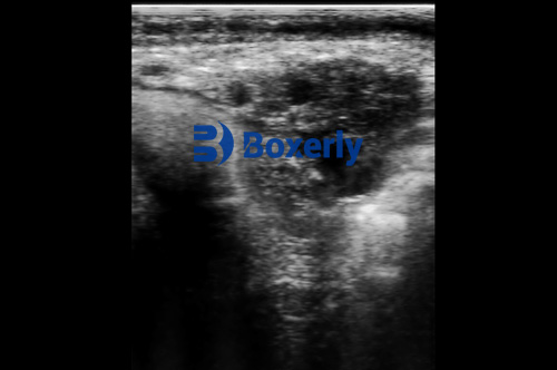
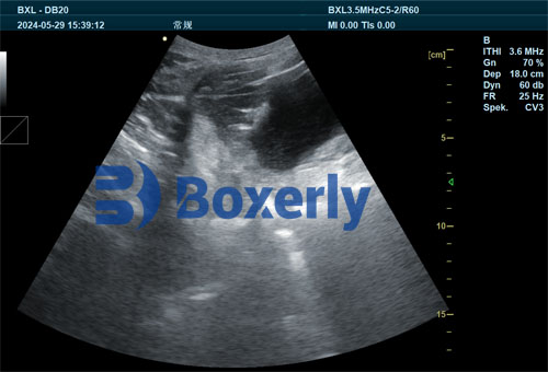
B-mode ultrasound offers two-dimensional cross-sectional imaging of the ovary, revealing follicles as hypoechoic (dark) spherical structures. High-frequency probes (7.5–10 MHz) provide better resolution, enabling detection of follicles as small as 1–2 millimeters in diameter.
For quantifying follicular reserve, veterinarians count the antral follicle count (AFC) visible on ultrasound. The AFC is a reliable proxy for the total primordial follicle pool, validated by correlation studies with histology and hormonal markers like anti-Müllerian hormone (AMH).
Standardized Scanning Protocols
To ensure accurate and repeatable follicle counts, a standardized scanning procedure is critical:
Restrain the animal calmly to minimize movement.
Use transrectal or transabdominal approaches depending on species and size.
Perform systematic scanning of the entire ovary in multiple planes to avoid missing follicles.
Record all follicles ≥2 mm, distinguishing sizes to evaluate follicle recruitment dynamics.
Emerging Technologies
Advancements include 3D ultrasound and Doppler imaging to assess ovarian blood flow, providing deeper insights into follicular health and viability. Though less common in routine farm practice, these technologies enhance precision in research and elite breeding programs.

International Perspectives on Ultrasound Quantification of Follicular Reserve
Globally, veterinary reproductive specialists recognize the value of ultrasound for managing breeding animals. Studies across Europe, North America, Australia, and parts of Asia demonstrate how AFC monitoring aids genetic selection and fertility prediction.
For instance, Australian cattle breeders use AFC to identify cows with superior fertility traits, incorporating this data into breeding value indices. In Europe, research on sheep has linked ultrasound-derived follicular counts to lambing rates and prolificacy, guiding selective breeding for increased flock productivity.
In pig breeding, ultrasound is used to monitor ovarian follicular dynamics during different reproductive phases, improving sow replacement decisions and litter size optimization.
Practical Applications and Benefits in Livestock Farming
Enhancing Breeding Management
Ultrasound quantification of follicular reserve enables:
Early identification of animals with low ovarian reserves, reducing costly breeding failures.
Tailoring of reproductive interventions, such as hormone treatments, to maximize ovulation rates.
Improved timing for artificial insemination or embryo transfer based on follicle status.
Selection of replacement stock with higher fertility potential, extending productive herd life.
Economic Impact
By avoiding repeated breeding attempts on animals with depleted reserves, farmers reduce feed, labor, and veterinary costs. Furthermore, maintaining a herd with robust ovarian reserves increases overall fertility, litter size, and offspring quality, directly boosting farm profitability.
Animal Welfare and Sustainability
Non-invasive ultrasound assessment minimizes stress and physical harm, aligning with ethical livestock management. Sustainable breeding programs supported by ultrasound data promote genetic diversity and long-term herd health.
Limitations and Challenges
While ultrasound is a powerful tool, some limitations exist:
Operator skill heavily influences image quality and follicle detection accuracy.
Small follicles under 1 mm remain difficult to detect, potentially underestimating reserve.
Variability between breeds and individuals necessitates population-specific reference values.
Equipment cost and maintenance may restrict use in smaller farms or developing regions.
Continued training and technological improvements aim to overcome these challenges.
Future Directions in Veterinary Ultrasound and Follicular Reserve Research
Emerging biomarkers combined with ultrasound imaging, such as AMH measurement, promise integrated fertility assessments. Artificial intelligence algorithms are being developed to automate follicle detection and counting, reducing operator bias and improving throughput.
Genomic studies linked with ultrasound data could accelerate genetic improvement by identifying molecular markers associated with ovarian reserve longevity.
Conclusion
Veterinary ultrasound technology has revolutionized the ability to quantify follicular reserve depletion in aging breeding animals. This non-invasive, real-time imaging approach empowers farmers, veterinarians, and researchers to make precise, data-driven decisions that optimize reproductive efficiency, economic returns, and animal welfare.
International experiences confirm the technique’s broad applicability across livestock species and production systems. While challenges remain, ongoing technological advances and growing awareness promise wider adoption.
In a world increasingly focused on sustainable agriculture and food security, leveraging ultrasound for follicular reserve monitoring represents a vital step toward smarter breeding management and healthier, more productive livestock populations.
References
Ireland, J.J., Smith, G.W., Scheetz, D., & Smith, M.F. (2020). Use of ovarian ultrasonography to assess follicular reserve and fertility potential in cattle. Theriogenology, 150, 31-41. https://doi.org/10.1016/j.theriogenology.2020.01.012
Abreu, C.A., et al. (2019). Anti-Müllerian hormone and antral follicle count as predictors of fertility in sheep. Reproduction in Domestic Animals, 54(2), 159-167. https://doi.org/10.1111/rda.13462
Australian Beef Research Council. (2022). “Antral Follicle Count for Breeding Herd Management.” https://beefresearch.org.au/antral-follicle-count
U.S. Department of Agriculture. (2023). “Ultrasound in Swine Reproductive Management.” https://www.usda.gov/swine-ultrasound-repro
tags:


