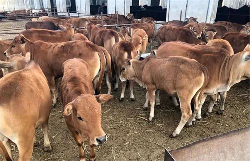
As any cattle farmer knows, the cow’s reproductive system is sensitive, complex, and often influenced by her overall energy status. Among the many tools used to assess reproductive health, ultrasound imaging of ovarian follicles stands out. When cows go through a period of negative energy balance (NEB)—typically during early lactation—things can get tricky. Even if a cow looks healthy from the outside, her ovarian activity might tell a different story.

Let’s talk about how follicle imaging reveals poor development during NEB, and why this matters so much for herd fertility and long-term productivity.
What Is Negative Energy Balance and Why It Matters
Negative energy balance happens when a cow uses more energy than she consumes. It’s especially common in high-yielding dairy cows right after calving. The cow’s body prioritizes milk production, often at the expense of other systems—like reproduction.
During NEB, fat reserves are mobilized, and blood metabolites like non-esterified fatty acids (NEFA) and ketones spike. These changes interfere with hormone secretion, leading to delayed or abnormal follicular development. The result? Fewer cows cycling naturally, poor conception rates, and more days open.
And that’s exactly where follicle imaging with ultrasound makes a huge difference.
What Follicle Ultrasound Tells Us About Reproductive Health
Using real-time B-mode ultrasound, we can visualize the cow’s ovaries, measure follicle size, track the growth of dominant follicles, and assess luteal structures. This is an essential, non-invasive way to monitor reproductive activity.
Here’s what we tend to observe during NEB:
Delayed emergence of dominant follicles: Follicles develop slowly, and often fail to reach the pre-ovulatory stage.
Smaller follicle size: Dominant follicles may stagnate below the typical 10–20 mm diameter.
Increased follicular atresia: Many follicles degenerate instead of maturing.
Low luteal tissue presence: Even when ovulation occurs, the resulting corpus luteum (CL) may be underdeveloped.
In practical terms, these cows are much less likely to conceive, even with timed artificial insemination.
What I’ve Seen on Farm: Real Imaging, Real Frustrations
In my own work with dairy herds, follicular imaging during the first 30 to 60 days postpartum often tells a discouraging story. We’ll scan cows that are producing well and eating decently—but when the probe goes in, the ovaries are quiet. No dominant follicles, no CL, just tiny structures that never fully mature.
In one herd, we followed 20 high-yield cows weekly after calving. By week 4, only 5 had developed a dominant follicle over 10 mm. The rest showed either arrested growth or multiple small, static follicles. None of them ovulated by week 6.
But the picture changed dramatically once feed intake increased and body condition improved. By week 8, nearly all cows resumed follicular activity. Imaging made it possible to track this rebound and adjust reproductive protocols accordingly.
Why NEB Disrupts Follicle Development
The science behind this is well documented. During NEB:
Low insulin and IGF-1 levels: Both are essential for follicular recruitment and growth.
High NEFA and beta-hydroxybutyrate: These impair granulosa cell function, leading to arrested follicular development.
Impaired LH pulsatility: Reduced luteinizing hormone release interferes with follicular maturation and ovulation.
Inflammatory cytokines: Often elevated during energy deficit, these disrupt ovarian cell signaling.
The combination of these metabolic shifts creates a hormonal environment that’s simply not conducive to reproduction.
Using Ultrasound to Guide Management Decisions
Follicle imaging isn’t just about diagnosis—it’s about decision-making.
If I scan a cow on day 30 postpartum and see no dominant follicle, that’s a red flag. She may need more time before insemination, or perhaps metabolic support through diet or supplements. On the other hand, if a large, healthy follicle is present with a good uterine tone, I can proceed with AI confidently.
Follicle imaging helps answer critical questions like:
Is this cow ready for breeding?
Is her body responding to synchronization protocols?
Do we need to recheck for ovarian cysts or silent heats?
Is it time to involve hormonal treatments?
This targeted approach avoids wasted semen, reduces frustration, and improves conception rates.
Nutrition Is Half the Battle
Even the best imaging protocol can’t compensate for poor nutrition. Reproductive function is tightly linked to the cow’s dietary energy intake, especially during early lactation.
Studies consistently show that cows with better dry matter intake and fewer body condition score losses post-calving resume estrous cycles sooner. That’s why I always pair follicular ultrasound data with feeding evaluations.
Some nutrition tips that help support healthy follicle development:
Maintain body condition around 3.25 at calving.
Encourage feed intake by offering high-quality forage and balanced energy.
Avoid excessive protein that leads to high urea levels, which can impair fertility.
Consider adding protected fats and rumen-stable supplements for energy density.
If energy input can match or slightly exceed energy output, follicular growth resumes, and reproductive success follows.
Follicular Cysts or Just Stalled Follicles?
One tricky area in imaging is telling the difference between a true follicular cyst and a large, non-ovulating follicle caused by NEB. Both appear as large, anechoic (black) structures on ultrasound—but they behave differently.
With a true cyst, the structure often persists for over 10 days and there’s no luteal tissue. In contrast, stalled follicles might regress or slowly develop over time. Repeat scanning is key to sorting this out.
Some vets use color Doppler to assess blood flow around the follicle wall. High vascularization may indicate a CL forming, while avascular follicles suggest poor development. This can be useful when you’re unsure what you’re looking at.
Rebuilding Reproductive Momentum
Once the cow moves past the NEB phase—usually around 60–70 days postpartum—follicular imaging often shows a resurgence in activity. Dominant follicles grow larger, CLs become more visible, and heats are easier to detect.
That’s when it’s time to re-evaluate your breeding timeline. Don’t rush it during NEB. Instead, use ultrasound to catch the moment when the cow is truly back in balance.
For many herds, waiting just 10–15 extra days before breeding can mean the difference between a 30% and a 60% conception rate.
Final Thoughts
Follicular imaging is more than a high-tech diagnostic tool—it’s a window into how a cow is really doing. During NEB, even when she’s eating and milking well, her ovaries might be struggling. And you can’t fix what you don’t see.
By combining real-time ultrasound with smart nutritional management and patient reproductive timing, farmers can drastically improve fertility outcomes.
Follicular development isn’t just a matter of biology—it’s a reflection of the entire cow’s energy status. Ultrasound shows us that, plain and simple.
tags: Cow Follicle Development


