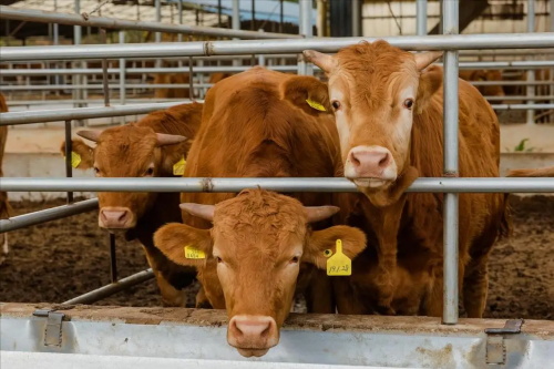
If you’re involved in cattle farming or veterinary medicine, you’ve probably heard about cystic ovary syndrome (COS) in cows. It’s a tricky condition that messes with the cow’s reproductive system and can cause all kinds of headaches, from delayed conception to reduced milk production. What’s fascinating is how much follicle growth in the ovaries reveals about this syndrome—and how ultrasound technology has become a game changer for diagnosing it.

Understanding Follicle Growth in Cows
First off, let’s talk about follicles. These are tiny sacs within the ovary that contain eggs. In a healthy cow, follicles develop in a cyclical pattern—growing, maturing, and then releasing an egg during ovulation. This cycle is critical for regular estrus (heat) and successful breeding.
Follicle growth happens in waves. Usually, two or three waves of follicles develop during a cow’s estrous cycle, with one dominant follicle eventually ovulating. The rest regress naturally. This follicular wave pattern is quite well understood thanks to decades of research and is pretty consistent across most breeds.
When everything’s working smoothly, a cow will show normal estrous behavior and conceive within a predictable timeframe. But if the follicle development gets disrupted, problems start creeping in.
What Happens in Cystic Ovary Syndrome?
Cystic ovary syndrome in cows is somewhat similar to the PCOS (polycystic ovary syndrome) that human females can get, though the causes and symptoms differ. In cows, COS is defined by the presence of ovarian cysts—fluid-filled sacs that can be quite large and don’t ovulate properly.
Instead of progressing through the typical follicular wave, a follicle can grow abnormally large but fail to release the egg. This results in a cyst that can linger for weeks, interfering with normal hormonal cycles. The cow may show irregular or absent estrus signs, making it tough to breed her on time.
The big issue here is that these cysts mess with the hormone feedback loops controlling reproduction, especially the levels of luteinizing hormone (LH) and follicle-stimulating hormone (FSH). Without proper ovulation, conception rates drop and the cow’s reproductive efficiency takes a hit.
How Follicle Growth Patterns Change with COS
Follicle growth in cows with cystic ovary syndrome tends to follow an abnormal trajectory. Instead of the normal rise and fall, the dominant follicle stalls in growth or becomes cystic. Sometimes multiple cysts can be present, further complicating the hormonal environment.
From what vets and researchers observe, cows with COS often have:
Prolonged follicular phases where follicles fail to ovulate
Irregular follicle size with cysts typically larger than 2.5 cm in diameter
Lack of corpus luteum formation since ovulation doesn’t happen
Hormonal imbalances detected via blood tests, matching follicle dysfunction
One of the reasons this syndrome is so challenging is that the signs can be subtle, especially in herd settings where individual cow behavior is harder to track. Cows might have mild or no obvious estrus signs, or they might display behavioral changes that look like other issues.
Ultrasound: The Best Friend for Detecting COS
This is where ultrasound shines—literally and figuratively. Over the past couple of decades, ultrasound has become the preferred tool for monitoring ovarian follicle development and diagnosing cystic ovary syndrome in cattle.
Before ultrasound, diagnosis relied mostly on rectal palpation and behavioral observation, which are less accurate and more subjective. Ultrasound offers a real-time, non-invasive way to visualize follicles, cysts, and the presence or absence of the corpus luteum.
Thanks to advances in portable ultrasound machines, vets can perform on-farm scans and get instant information. This allows early detection of COS, so interventions like hormonal therapy or management changes can start sooner.
How Ultrasound Helps Track Follicle Growth
During an ultrasound exam, the veterinarian scans the cow’s ovaries via rectal approach, capturing images of follicles and cysts. Follicles appear as fluid-filled, round structures on the ovary’s surface.
By measuring the size and number of follicles, vets can assess whether the cow’s ovarian activity is normal or abnormal. For instance:
Follicles smaller than 1 cm are usually in early growth stages
Dominant follicles range from about 1.5 to 2.5 cm and are expected to ovulate
Follicles larger than 2.5 cm persisting beyond 10 days are suspicious for cysts
With repeated scans, vets can observe whether follicles regress or persist abnormally. This temporal information is crucial because a single scan might not capture the whole picture.
Moreover, ultrasound imaging can detect the absence of corpus luteum—a sign that ovulation has not taken place. This detail helps differentiate cystic follicles from normal structures.
What Foreign Research and Practices Say
In the US, Europe, and Australia, ultrasound-based diagnosis of COS in cows is widely accepted and has become part of standard reproductive management in many farms. Research papers highlight the strong correlation between follicular dynamics observed via ultrasound and successful treatment outcomes.
For example, studies from the University of Wisconsin and University of Sydney emphasize that:
Early identification of cysts through ultrasound significantly improves pregnancy rates post-treatment.
Ultrasound-guided classification of cysts (follicular vs luteal cysts) helps tailor hormonal treatments better.
Regular ultrasound monitoring can reduce unnecessary culling of cows that might recover with proper intervention.
Farmers familiar with these findings often prioritize ultrasound exams, especially during transition periods postpartum when cows are more susceptible to reproductive disorders.
Practical Tips for Farmers and Veterinarians
If you’re dealing with COS on your farm or in your practice, here are some useful takeaways based on international experience:
Routine Ultrasound Scanning: Implement regular ovarian scanning during the postpartum period (30-60 days after calving) when cows are most vulnerable to cyst formation. Early detection means quicker treatment.
Observe Follicle Size and Persistence: Watch for follicles that don’t regress after 10 days and grow larger than 2.5 cm. These should raise suspicion for cysts.
Combine with Hormone Testing: When possible, complement ultrasound findings with progesterone blood tests to confirm ovulation status and ovarian function.
Use Targeted Hormonal Treatments: Based on ultrasound diagnosis, treatments with GnRH or prostaglandins can be more effectively timed to resolve cysts.
Record Keeping: Maintain detailed records of follicle growth patterns and treatment outcomes to improve herd reproductive performance over time.
Why Follicle Growth Matters Beyond COS
It’s not just about cysts. Monitoring follicle growth is also essential for optimizing breeding programs. Knowing when a cow’s dominant follicle is ready allows for better-timed artificial insemination or natural breeding.
Plus, abnormal follicle dynamics can signal other underlying health or metabolic issues, like energy imbalance or stress. So, ultrasound scans provide a window into the cow’s overall reproductive health.
In many countries, veterinary extension services encourage farmers to adopt ultrasound scanning not only for diagnosing cystic ovaries but also as part of a broader reproductive health monitoring program.
The Future of Follicle Growth Monitoring and COS Diagnosis
With ongoing technological advances, ultrasound devices are becoming more affordable, portable, and user-friendly. There’s also growing interest in integrating artificial intelligence to analyze ultrasound images for quicker and more accurate follicle evaluation.
Moreover, researchers are exploring new biomarkers alongside follicle growth patterns to predict COS risk before cysts develop, potentially allowing preventive strategies.
In short, the link between cow follicle growth and cystic ovary syndrome diagnosis will remain a hot topic, evolving with new tools and better understanding.
tags: Cow Follicle Development


