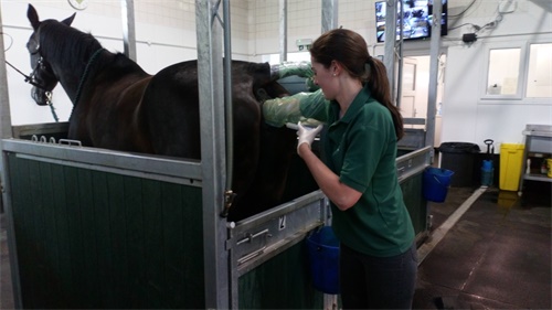
For horse breeders, the final trimester of pregnancy is a time of excitement, close monitoring, and cautious decision-making. At this stage, the health of both the mare and the unborn foal is paramount. While visual signs like abdominal enlargement and behavioral changes give some indication of late pregnancy progress, nothing compares to the accuracy and reassurance that ultrasound imaging provides.

Across breeding farms worldwide—from Kentucky to New South Wales—ultrasound has become a cornerstone of equine reproductive management, particularly during the final stages of gestation. The ability to “see” inside the mare’s uterus gives veterinarians and breeders the confidence to act early if complications arise, and to ensure the foal is developing normally. Let’s take a deeper look at how late-pregnancy ultrasound is used in daily practice and why it’s such a vital part of successful horse breeding.
What Makes Late Pregnancy So Critical?
Late pregnancy in mares typically begins around day 210 of gestation. Since the average equine pregnancy lasts around 340 days, the final three months are marked by rapid fetal growth and shifting metabolic demands on the mare. It’s during this phase that most fetal abnormalities—such as abnormal positioning or restricted growth—become detectable.
At this stage, veterinary professionals rely heavily on transabdominal and transrectal ultrasound to track changes in fetal position, heartbeat, size, and fluid levels. Unlike early pregnancy, where the focus is often just on confirming pregnancy and checking for twins, late pregnancy calls for more detailed and repeated assessments.
A Window Into the Uterus: What Ultrasound Reveals
Horse breeders often describe ultrasound as a “third eye” on the farm. It gives a direct view into the uterus and reveals critical details that aren’t detectable with palpation or external observation.
Fetal Position and Movement
Fetal positioning is a top concern during the final trimester. Mares are most likely to have a successful birth if the foal is in anterior presentation (head and forelimbs first). If the fetus is in breech or transverse position, the risk of dystocia (difficult birth) increases dramatically. Regular ultrasounds help identify malpositioning early enough to take preventive steps.
Fetal Heart Rate
An ultrasound allows veterinarians to assess the fetal heart rate—an essential indicator of fetal well-being. A normal fetal heart rate ranges from 65 to 115 bpm during late pregnancy. Deviations can signal stress, hypoxia, or even placental separation. In such cases, immediate intervention might be required, such as initiating treatment or planning an early delivery.
Placental Health and Thickness
Ultrasound also helps measure the combined thickness of the uterus and placenta (CTUP), particularly in the cervical star region. Abnormally thickened CTUP can be a sign of placentitis—an infection that can result in premature birth or fetal loss if left untreated. The ability to detect placental pathology through ultrasound allows for timely antibiotic or anti-inflammatory therapy.
Amniotic and Allantoic Fluid Volume
The quantity and quality of uterine fluids are critical for fetal survival. An ultrasound can assess whether fluid levels are within the normal range and check for signs of meconium, debris, or infection. Reduced fluid or abnormal content can suggest compromised fetal health.
How Often Do Breeders Scan Late in Pregnancy?
There’s no strict universal schedule, but many experienced breeders follow a scanning protocol that includes:
Initial third-trimester check: Around day 220–240 to assess fetal growth and placental thickness
Mid-trimester recheck: Around day 270 to evaluate fetal position and heartbeat
Final pre-foaling scan: Between days 300–320 to confirm normal development and estimate delivery readiness
Veterinarians may adjust the frequency depending on breed, the mare’s history (especially if she’s had previous abortions), or any complications that arise. For example, mares with placentitis risk might be scanned every 7–10 days until delivery.
Tools and Techniques: What’s Used on Farms?
Most farms use portable B-mode ultrasound systems with 3.5–5 MHz convex or microconvex probes for abdominal scans. For transrectal scans—still common in the early and mid-stages of pregnancy—a 5–7.5 MHz linear probe is preferred. In the late stage, abdominal access becomes more important due to fetal descent and uterine positioning.
The best machines for this job aren’t necessarily the most high-end. What matters more is image clarity, durability, and ease of operation in barn environments. Many farms opt for veterinary-dedicated models that offer freeze-frame capabilities, video loop storage, and easy cleaning.
Interpreting Findings: Experience Matters
Reading late pregnancy ultrasound images isn’t always straightforward. For example, fetal movement may be limited in large mares, and distinguishing between amniotic and allantoic fluids takes skill. That’s why breeding farms often work closely with veterinarians who specialize in equine reproduction.
One common challenge is interpreting CTUP measurements. While a reading above 12–13 mm may indicate placentitis, individual variation exists, especially between draft breeds and lighter mares. Context is key: a single abnormal reading doesn’t always signal danger, but a rising trend over multiple scans usually requires action.
Real-World Benefits: Why Farms Rely on Ultrasound
Veterinarians and breeders alike praise ultrasound not just for its accuracy, but for the peace of mind it provides. Here’s how it helps in real scenarios:
Preventing Sudden Losses: Early detection of placental problems or abnormal fluid lets the vet treat the mare before fetal distress escalates.
Planning Foaling Timing: By assessing fetal maturity (e.g., lung echogenicity, ossification), ultrasound can help estimate when a foal is likely to arrive and reduce the guesswork around foaling time.
Monitoring High-Risk Mares: Older mares, those with prior complications, or those carrying IVF embryos are under stricter surveillance, where ultrasound is the best monitoring tool available.
Supporting Foal Viability: In some cases, farms schedule pre-foaling exams to prepare for high-risk deliveries with advanced neonatal care on standby.
A Tool That’s Here to Stay
Equine reproduction is both science and art. Ultrasound has bridged the gap between guesswork and precision. It's not just about spotting trouble—it’s about staying one step ahead of it. On large breeding operations, this tool has become just as essential as a halter or foaling alarm.
The beauty of ultrasound lies in its non-invasiveness. Mares tolerate scans well, and foals remain undisturbed in the process. Unlike blood tests or biopsy methods, there’s no need for sedation, restraint, or recovery time. It’s efficient, safe, and repeatable.
Looking Ahead: Evolving Applications
As imaging technology improves, breeders are starting to use ultrasound for more than just monitoring. Some are now measuring fetal limb length and organ size to predict foal maturity. Others are experimenting with Doppler imaging to get even more data on fetal circulation.
Meanwhile, portable, battery-powered units are making it easier than ever to scan in the field, even without a clinic or power source. This accessibility has opened the door for small farms to adopt best practices once limited to elite operations.
Final Thoughts
What was once a luxury is now a routine part of breeding management. Ultrasound during late pregnancy has reshaped how horse breeders approach the final stretch of gestation. It turns anxious waiting into proactive care and replaces worry with actionable insight.
Whether you’re managing a Thoroughbred facility or raising warmbloods on a small farm, knowing what’s going on inside the mare during late pregnancy can make all the difference. A healthy foal starts with informed care—and in today’s world, that often means having an ultrasound probe in hand.
tags:
Text link:https://www.bxlultrasound.com/ns/866.html


