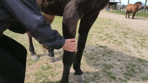
When it comes to keeping horses healthy and sound, tendon injuries are one of the biggest concerns for farmers, breeders, and equine veterinarians. These injuries can sideline a horse for months, and if not caught early, they can turn into chronic problems that end a horse’s working life entirely. That’s where ultrasound comes into play. Across farms in Europe, the US, Australia, and beyond, equine ultrasound has become a trusted tool for early tendon injury detection—saving both time and animals.
Why Tendon Health Is a Big Deal on Horse Farms
Tendons are the connective tissues that anchor muscle to bone. In horses, the superficial and deep digital flexor tendons, along with the suspensory ligament, are especially vulnerable. These tissues carry a heavy load during exercise, whether it’s a dressage routine, a gallop across the field, or simply trotting around a paddock. Even minor overuse or improper footing can lead to micro-tears or inflammation.
For working horses—especially racehorses, jumpers, or ranch horses—tendon injuries are often career-ending. That’s why farms have become more proactive in their approach. Rather than waiting for a horse to show obvious signs of lameness or swelling, many now use ultrasound to detect subtle changes in tendon structure before things get worse.
The Role of Ultrasound in Early Detection
Veterinary ultrasound works by sending sound waves into the body and capturing echoes that bounce back. This creates a real-time, black-and-white image of soft tissues like tendons. On a healthy horse, tendons appear as fine, parallel lines with uniform texture. But when there’s inflammation, fluid buildup, or fiber disruption, these changes show up clearly on the screen.
Farm managers and vets typically use a high-frequency linear transducer—often around 7.5 to 12 MHz—for the best image resolution. The exam is done standing, with the horse sedated or calmly restrained, and the leg area clipped and gelled to ensure a clear contact between the probe and the skin.
The beauty of this method? It’s non-invasive, quick, and can be repeated as often as needed without harming the horse.
What Farmers Are Looking For in the Scan
Experienced veterinarians and farm staff are trained to look for key indicators:
Loss of fiber pattern: Indicates damaged tendon fibers
Anechoic or hypoechoic areas: These dark regions signal fluid or inflammation
Tendon enlargement: A swollen tendon is often the first external clue
Changes in cross-sectional area (CSA): Increased CSA often means early-stage injury
Increased vascularization: With Doppler ultrasound, extra blood flow can signal healing—or active inflammation
Using ultrasound, even a subtle 10% increase in tendon diameter can be flagged, allowing time for rest and treatment before a full-blown injury sets in.
Ultrasound Use Isn’t Just for Diagnosis—It’s for Prevention Too
What’s truly changed in recent years is how farms are using ultrasound not just for diagnosis, but for routine monitoring. On high-performance farms, especially in Europe and the US, it’s common practice to scan horses every few weeks during training. This proactive approach helps spot microdamage before it becomes a tear.
Many horse farms now keep ultrasound health records, tracking each horse’s tendon status over time. That historical data is invaluable. If a tendon shows early warning signs, the trainer can adjust the horse’s workload, footing, or even shoeing to reduce strain.

Real-Life Example: A Stud Farm in Kentucky
At a large Thoroughbred farm in Lexington, Kentucky, ultrasound has become part of every horse’s conditioning program. Their veterinarians routinely scan yearlings as they begin light training. During one spring season, the team spotted a hypoechoic area in a colt’s superficial digital flexor tendon—he showed no signs of pain, but the scan revealed trouble brewing.
They rested the colt for four weeks, adjusted his footing, and followed up with another scan before returning him to training. That horse went on to win races at age three—an outcome that would’ve been unlikely had the injury progressed undetected.
Challenges and Limitations
Of course, ultrasound isn’t magic. It requires skill to interpret correctly, and not every tendon change leads to lameness. Overdiagnosis can happen, which is why having an experienced vet is key. Also, some deeper tendons, like those in the hind limb or pelvic region, are harder to visualize with standard probes.
Another challenge is cost. While ultrasound machines have become more affordable, they’re still an investment—especially if multiple units are needed across a large operation. However, many farms justify the cost by considering the long-term savings in vet bills, rehab costs, and lost training time.
Combining Ultrasound with Other Tools
Many farms now take a multi-modal approach: pairing ultrasound with lameness evaluations, thermography, or even motion analysis apps. If a scan picks up subtle tendon irregularities, trainers can compare that with gait analysis to see if there’s an underlying issue affecting the horse’s stride or load distribution.
Blood markers are also emerging as a future diagnostic companion. In some research settings, blood tests can pick up biomarkers associated with tendon inflammation. Combined with ultrasound imaging, the accuracy of early detection is getting stronger every year.
Ultrasound as a Communication Tool
One often overlooked benefit is how ultrasound bridges the gap between vet and owner. A good scan image provides a visual story—one that’s easy to understand. Farm managers can see exactly what’s happening in the tendon, making it easier to agree on next steps. This clarity builds trust and improves care outcomes.
For breeding farms, ultrasound also plays a role in evaluating tendon integrity in stallions and broodmares, helping determine whether they’re fit for work, riding, or pasture breeding. For older horses, it helps manage age-related tendon degeneration.
What's Next: The Future of Equine Ultrasound on Farms
Portable ultrasound units are already common on farms, but the future is looking even more high-tech. Some emerging tools include:
AI-powered image interpretation to reduce reliance on manual diagnosis
Cloud-based ultrasound record systems for shared monitoring between farm and vet
Wireless probes that send images directly to a phone or tablet
3D ultrasound scanning for more detailed tendon structure visualization
These technologies are likely to make tendon monitoring more precise, accessible, and consistent across farms of all sizes.
Conclusion
On horse farms around the world, ultrasound has quietly become one of the most effective tools in preventing and managing tendon injuries. It’s non-invasive, real-time, and powerful—not just for diagnosis, but for keeping horses sound before problems even begin. Early detection saves time, money, and most importantly, keeps our horses healthier and happier.
Whether it’s a Kentucky racehorse, an Australian polo pony, or a British eventer, the message is the same: tendons matter, and ultrasound helps protect them.
tags:
Text link:https://www.bxlultrasound.com/ns/865.html


