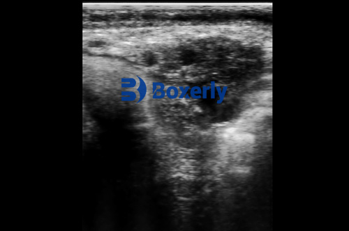
Subclinical mastitis is one of those silent threats on dairy farms—it doesn’t show up with obvious signs like clots in the milk or swelling in the udder, but it can quietly chip away at milk yield and animal welfare. Over the years, many dairy producers have relied on somatic cell counts (SCC) or California Mastitis Tests (CMT) to identify it. But what if there was a faster, more visual, and less subjective way to detect it—right in the barn? That’s where portable ultrasound technology steps in, and it’s changing how we catch mastitis before it becomes a bigger issue.

What Makes Subclinical Mastitis So Tricky?
Most farmers are pretty tuned in when a cow gets sick. But subclinical mastitis flies under the radar. The cow keeps eating, keeps milking, and doesn’t show pain, but there’s inflammation brewing in the udder. Over time, that can lead to tissue damage, lower milk quality, and a higher risk of chronic infection.
The tough part is that by the time SCC levels get high enough to trigger alarms, the cow may already be losing productivity. That’s why early detection is so valuable—and ultrasound is giving us a new way to spot the early changes in udder tissue that SCC tests can’t show.
What Can Ultrasound Actually See in the Udder?
Using a farm-ready portable ultrasound scanner, vets and trained farmers can look directly into the udder tissue and milk cisterns. Unlike palpation or lab testing, this gives you real-time visual information. Here's what we can pick up on an ultrasound image:
Echogenicity of mammary tissue: Healthy udder tissue has a certain "grain" on the screen. When inflammation starts, this texture changes. Areas might appear more hyperechoic (brighter) due to fibrotic development or hypoechoic (darker) with fluid build-up.
Duct and cistern dilation: Inflammation can cause enlargement or irregular shapes in milk ducts and cisterns—visible signs that something's off.
Localized fluid or abscess pockets: These are rare in subclinical cases but might appear in cows with repeated infections that never quite clear up.
With regular scanning, farms can monitor these changes over time and even compare images from different quarters. That’s huge when you’re managing large herds and want to spot issues early.
How Portable Ultrasound Fits Into Daily Dairy Work
Let’s face it—no farmer has time to haul cows to a vet clinic for every little checkup. That’s why portable ultrasound units are such a game changer. They’re built to be tough, waterproof, and battery-powered for long field use.
With models like the BXL-V50, you don’t need a specialized imaging room or even a power outlet. Just apply the gel, place the probe on the udder, and the screen shows you everything inside. These scanners often have freeze-frame, zoom, and image save features too, making it easy to share findings with your vet or save for herd health records.

Advantages Over Traditional Mastitis Testing
A lot of producers are used to SCC-based detection and might wonder—why switch it up?
Here’s where ultrasound offers added value:
Visual confirmation: Instead of relying on indirect indicators, you can actually see what’s happening inside the udder.
Quarter-specific accuracy: SCC often reflects whole-animal health, but ultrasound lets you assess individual quarters, catching early signs in one gland before it spreads.
Non-invasive and cow-friendly: No need to extract milk or cause stress. A few seconds of contact with a probe is all it takes.
Helps with treatment decisions: If a cow’s udder tissue is already showing chronic damage, it might not respond well to antibiotics. Ultrasound helps prioritize which cows to treat and which to cull.
Monitoring recovery: After a case of mastitis, follow-up ultrasound scans can track tissue healing and help you decide when it’s safe to return the cow to the milk line.
Real Experiences from the Field
Dairy producers in the U.S., Europe, and Australia are starting to use ultrasound not just for pregnancy checks but also for udder health. Take Mark, a Wisconsin dairy farmer managing 220 Holsteins. He started using ultrasound after multiple cows developed repeat mastitis issues post-treatment.
“We’d treat them, SCC would drop a little, then shoot back up. Ultrasound helped us spot which quarters had scarring that wasn’t going to heal. It helped us make the tough decisions faster,” he said.
Or consider Emma, a herd manager in the UK who uses the BXL-V50 scanner twice a month. “We don’t just wait for SCC spikes anymore. Now we do spot checks on high-producing cows and early lactation animals. It’s added maybe 15 minutes to the routine, but saved us thousands in treatment costs.”
When and How to Scan
Ultrasound isn’t meant to replace all other testing—but it complements it beautifully. The best time to scan for subclinical mastitis is:
Early in lactation: Cows are most vulnerable to infection right after calving.
After clinical mastitis treatment: To evaluate healing and avoid reinfection.
Before dry-off: To identify cows that might benefit from a longer dry period or a different dry cow therapy.
As for technique, scanning is typically done with a convex or linear probe, depending on the cow’s udder shape and size. Most portable scanners are preset with suitable depth and contrast settings, but some models allow adjustment if needed. The probe is applied gently to the cleaned udder surface with gel, and the image appears instantly on the screen.
Where the Technology Is Headed
Veterinary ultrasound has come a long way from the early bulky machines of the 1990s. Modern portable models are lighter, clearer, and smarter. Some units now come with wireless capabilities to send images to a vet’s phone. Others integrate AI to flag suspicious tissue patterns automatically—though most experts still prefer the experienced eye of a vet or trained technician.
In the future, we’re likely to see even more automation and image analysis tools, maybe even mastitis scoring systems built into the scanner. But for now, even basic grayscale imaging offers a powerful edge.

Final Thoughts
For years, subclinical mastitis detection relied on lab work and hunches. Now, portable ultrasound is making it easier, faster, and more accurate. It’s not just a tool for veterinarians anymore—it’s a practical addition to the modern dairy producer’s toolkit.
By catching udder inflammation before it becomes obvious, farms can protect milk quality, reduce unnecessary antibiotic use, and keep cows healthier longer. And with tools like the BXL-V50 becoming more affordable and rugged, this isn’t high-end tech for elite operations—it’s everyday gear for everyday farms.
Sources & References
Harmon, R.J. (2020). Physiology of Mastitis and Factors Affecting Somatic Cell Counts. https://www.ncbi.nlm.nih.gov/pmc/articles/PMC10134552
Blowey, R.W., & Edmondson, P. (2010). Mastitis Control in Dairy Herds. CAB International.
University of Wisconsin Extension. (2023). Ultrasound Applications in Dairy Cow Health. https://fyi.extension.wisc.edu
tags:
Text link:https://www.bxlultrasound.com/ns/860.html


