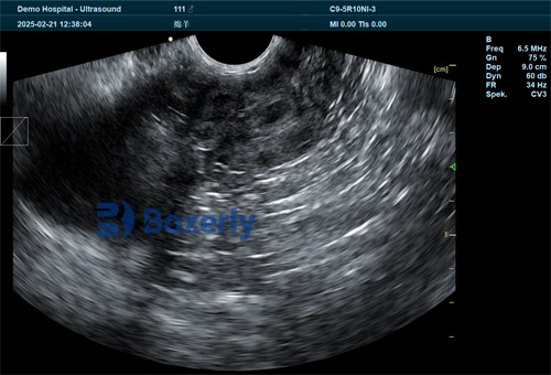
Among reproductive technologies used in sheep farming today, veterinary ultrasonography stands out as one of the most accessible, cost-effective, and non-invasive tools for reproductive management. For producers aiming to optimize breeding outcomes, improve flock productivity, and reduce feed and labor waste, ultrasound imaging provides real-time, reliable data on pregnancy status. However, understanding how ultrasound detection efficiency varies at different reproductive stages is key to making it truly useful.

In this article, we explore how sheep ultrasound examinations perform across three defined stages of early gestation. Drawing on both local practice and international understanding, we compare detection standards and effects from day 19 to day 50 post-mating, and identify the best time to conduct scanning in order to balance accuracy and practicality.
Understanding the Reproductive Timeline in Ewes
After successful mating or artificial insemination, a ewe's pregnancy progresses through several defined stages. Based on international research and field observations, we can divide early pregnancy ultrasound assessments into three practical stages:
Gestational Sac and Yolk Sac Stage (19–30 days post-mating)
Pregnancy Cavity Stage (25–40 days post-mating)
Placenta and Fetal Development Stage (35–50 days post-mating)
Each stage has its own ultrasound features and diagnostic limitations. Below we analyze each one in detail.
Ultrasound at the Gestational Sac and Yolk Sac Stage (19–30 Days Post-Mating)
In theory, skilled operators using high-resolution ultrasound equipment can detect early signs of pregnancy as early as 15–18 days post-mating. However, international and Chinese studies alike show that the diagnostic reliability during this period is low. The images are vague, and false negatives are frequent due to small sac sizes or operator inexperience.
For this reason, most veterinarians and researchers recommend starting pregnancy scanning from day 19 or 20 post-mating. At this point, detection accuracy improves significantly. Between days 20–22, the detection rate shows its most rapid increase. From day 23 to 26, the rate continues to rise but at a slower pace, peaking at day 24 with a detection success rate of around 81.25%.
Ultrasound images during this stage typically show a gestational sac first (day 19–22), followed by clearer yolk sac visualization (day 22–28). Both structures are best observed between days 24–26. Beyond this window, the yolk sac begins to shrink and becomes harder to detect, while the gestational sac gets gradually replaced by the developing pregnancy cavity.
Ultrasound at the Pregnancy Cavity Stage (25–40 Days Post-Mating)
This stage marks a transition in the visible uterine contents. By around day 26, the developing pregnancy cavity begins to dominate the image. Detection rates rise sharply from day 27 to 31—the primary growth phase—and continue at a slower pace from day 32 to 37.
By day 32, diagnostic accuracy reaches a peak of about 93.75%. From an international perspective, this matches well with findings from sheep-rearing regions in Australia, New Zealand, and the UK, where ultrasound at this stage is often used for both pregnancy confirmation and fetal number estimation.
Between days 32–37, the pregnancy cavity is at its most distinguishable. However, after day 37, a new pattern emerges. The visualization of placental and fetal structures increases rapidly, making pregnancy cavity signs less distinct. In practice, this means that while detection is still possible, the focus of the scan shifts toward fetal viability and development rather than simple confirmation.
Ultrasound at the Placenta and Fetal Stage (35–50 Days Post-Mating)
Starting around day 35, ultrasound scans can begin detecting clear fetal structures and placental zones. Between days 36–40, the detection rate of these features increases dramatically. A secondary increase follows between days 40–43, with maximum detection accuracy (near 100%) achieved by day 42.
Interestingly, the international veterinary literature supports this timeline. Studies in North America and Europe recommend scanning between 35–45 days post-mating for maximum fetal clarity and fetal number determination. This window also aligns with embryo transfer programs that rely on early fetal health assessment.
During days 40–43, placental visualization dominates, with fetal structures becoming more prominent after day 43. Heartbeats, limb movements, and cranial formations are often visible during this stage, offering not just confirmation of pregnancy, but also information on fetal viability.
Comparative Analysis of Detection Effectiveness
To determine which scanning window offers the best trade-off between accuracy, convenience, and diagnostic benefit, we compare the three stages using T-tests and field results. While detection rates at day 24 (81.25%), day 32 (93.75%), and day 37 (100%) all meet production needs, the differences between them are not statistically significant (P > 0.05).
However, considering the operational logistics—such as scheduling, labor, and ewe handling—the optimal scan time is around day 32. At this point:
Detection is highly accurate
The pregnancy cavity is clearly visible
Yolk sac or placental confusion is minimal
Ewes are far enough along to rule out early embryonic loss
Management decisions (e.g., culling non-pregnant ewes, adjusting nutrition) can be made efficiently
Therefore, while earlier or later scans can still be justified, day 32 emerges as the most efficient window for herd-level management.

Global Perspectives on Sheep Ultrasonography
In sheep-producing countries such as Australia, Ireland, and the U.S., veterinary ultrasonography is widely adopted and seen as essential for modern flock reproductive management. Portable, durable ultrasound machines allow producers to scan in-field, reducing stress to animals and minimizing disruption to feeding or lambing routines.
Additionally, foreign studies emphasize training as a major factor in scan success. For example, in New Zealand, where scanning services are often outsourced to trained technicians, the average detection rate across farms is consistently over 95%, especially during the 30–40 day window.
The growing availability of real-time B-mode ultrasonography has also opened up possibilities beyond pregnancy confirmation—such as early sexing of fetuses, detection of twins, and even diagnosing reproductive pathologies like endometritis.

Conclusion
Ultrasound technology has revolutionized pregnancy diagnosis in sheep. By understanding how detection patterns vary across early gestation stages—from the yolk sac to placental formation—producers can make more informed decisions on when and how to scan their ewes.
While reliable pregnancy detection can begin around day 19, the optimal window that balances high accuracy and on-farm practicality is around day 32 post-mating. This timing maximizes both detection performance and economic return, reducing the risk of unnecessary culling or feeding non-pregnant animals.
As global sheep production continues to modernize, veterinary ultrasonography will remain a cornerstone of efficient reproductive management—bringing clarity, confidence, and cost-effectiveness to shepherds everywhere.
tags:
Text link:https://www.bxlultrasound.com/ns/820.html


