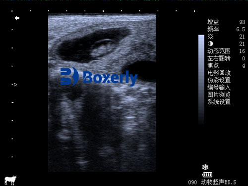
For dairy farmers, veterinarians, and researchers, understanding fetal development and early sex determination in pregnant cows has profound implications on herd management, breeding decisions, and economic planning. In recent decades, veterinary ultrasonography has emerged as a transformative, non-invasive tool that allows real-time imaging of bovine embryos and fetuses in utero. Especially between gestation days 55 and 75, ultrasound enables accurate estimation of fetal age, monitoring of organ growth, identification of developmental abnormalities, and determination of fetal sex. In this article, I will explore how ultrasound is applied in embryological imaging and fetal sexing in dairy cattle, supported by both research literature and practical use cases from international dairy systems.

Understanding Fetal Development Through Veterinary ultrasound
Veterinary ultrasound, specifically B-mode (brightness mode) imaging, has revolutionized reproductive management in cattle. Unlike traditional rectal palpation, ultrasound provides a visual representation of the developing embryo or fetus, enabling early pregnancy confirmation and allowing practitioners to monitor embryonic structures in detail.
In bovine fetuses, key anatomical landmarks—such as the head, limbs, eyes, stomach, spine, and ribs—can be visualized as early as 30–40 days post-conception. As the pregnancy progresses, skilled operators adjust the angle and depth of the transrectal probe to capture the clearest image slices of fetal organs. These measurements are then used to estimate gestational age and predict parturition timing, which is crucial for optimizing labor, nutrition, and calving support schedules.
In many countries such as the United States, Canada, and Australia, ultrasound scanning is incorporated as a routine check in breeding herds. Research published by the University of Wisconsin (Pohler et al., 2020) demonstrates that the size of fetal organs, such as biparietal diameter (head width), crown-rump length, and ocular diameter, are strongly correlated with gestational age. Veterinarians use these parameters to establish growth curves for healthy fetal development and to detect deviations that may signal intrauterine growth retardation (IUGR) or early embryonic loss.
Practical Advantages on the Farm
For dairy producers, accurate fetal aging using ultrasound helps to:
Reduce calving complications by predicting delivery dates
Improve dry-off timing and adjust nutritional plans
Detect twin pregnancies early to provide additional care
Identify developmental problems like uterine infections or abnormal fetal positioning
Moreover, building a reference library of ultrasonographic fetal images at different gestational stages has proven valuable for veterinary training and herd reproductive audits. For example, veterinary schools in Europe and the U.S. now offer specialized modules in bovine embryology imaging to train students in interpreting these diagnostic images.
Ultrasound-Based Fetal Sex Determination
One of the most valuable applications of veterinary ultrasound in mid-gestation dairy cows is fetal sex determination. Typically conducted between days 55 to 75 of gestation—with day 67 being optimal—this technique helps producers make informed decisions regarding heifer retention, marketing strategies, and genetic planning.
The process hinges on locating the genital tubercle, a small embryonic structure that later develops into male or female external genitalia. Around day 55, the genital tubercle migrates differently in male and female fetuses:
In males, it migrates toward the umbilical cord, so an additional bright spot (tubercle echo) appears near the cord region on the ultrasound image.
In females, it migrates toward the tail, so bright echoes appear closer to the tail base, often presenting as two or three reflective points.
By systematically scanning from the fetus’s abdomen to tail base, practitioners can distinguish sex-related echogenic points. Accuracy is typically over 95% in ideal conditions, although difficulties may arise if the fetus is too deep, curled in a tight position, or obscured by the cow’s intestinal gas or vigorous straining.
Best Practices in Field Application
Performing fetal sex determination with high accuracy requires several field-tested practices:
Proper restraint of the cow to minimize stress and movement
Use of high-frequency linear probes (5–7.5 MHz) for optimal resolution
Experience in differentiating between anatomical structures on real-time images
Sequential scanning of all fetuses in case of twins or multiple gestations
After determining the sex of one fetus, practitioners should always continue scanning to check for the presence of additional fetuses and assess their position and sex. In countries like New Zealand and Germany, these protocols are standard in large-scale dairy operations, and many farms maintain digital ultrasound records for each pregnancy to track developmental trends.
Global Perspectives and Research
Globally, the integration of ultrasound into reproductive management has varied depending on veterinary service access, cost of equipment, and technician training. In North America and Northern Europe, most dairy farms routinely use ultrasound in herd reproductive programs. A study by De Vries and McGowan (2021) in Australia showed that routine fetal sexing increased the net profit per pregnancy due to more effective replacement heifer planning and calf management.
Furthermore, international research continues to refine imaging techniques. For instance, Canadian researchers are exploring the use of 3D ultrasound and Doppler imaging to assess blood flow in the umbilical cord, placenta, and fetal heart—opening possibilities for early detection of circulatory or developmental disorders. Meanwhile, in China and Brazil, collaborative projects are under way to develop AI-assisted fetal sex prediction based on stored ultrasound images, improving both speed and accuracy.

Challenges and Limitations
Despite its many benefits, ultrasound-based embryological imaging and sex determination still face some limitations:
Accuracy is reduced outside the optimal window of 55–75 days
Requires skilled operators to differentiate complex fetal structures
Equipment can be expensive for small-scale farmers
Certain anatomical anomalies or uterine positions can obscure imaging
Nevertheless, these challenges are increasingly being addressed through improved probe design, affordable portable ultrasound units, and standardized technician certification programs.
Conclusion
Ultrasound technology has become a cornerstone of modern dairy herd reproductive management. Its ability to visualize fetal development and determine fetal sex early in gestation allows dairy producers to optimize production strategies, improve animal welfare, and increase economic efficiency. From adjusting calving schedules to making data-driven decisions on replacement heifers, veterinary ultrasound imaging offers an invaluable window into the womb.
As global demand for precision livestock management grows, the role of ultrasonography will only become more significant. With ongoing advances in imaging resolution, software analysis, and AI integration, ultrasound imaging in dairy cattle promises to become even more accurate, accessible, and essential for farms around the world.
Reference Sources:
Pohler, K. G., et al. (2020). “Ultrasound use in bovine reproduction.” Journal of Dairy Science.
De Vries, M. J., & McGowan, M. (2021). “Economic benefit of fetal sex determination in dairy herds.” Australian Veterinary Journal.
American Association of Bovine Practitioners (AABP). (2023). “Guidelines for Bovine Ultrasonography in Reproductive Health.”
tags:
Text link:https://www.bxlultrasound.com/ns/819.html


