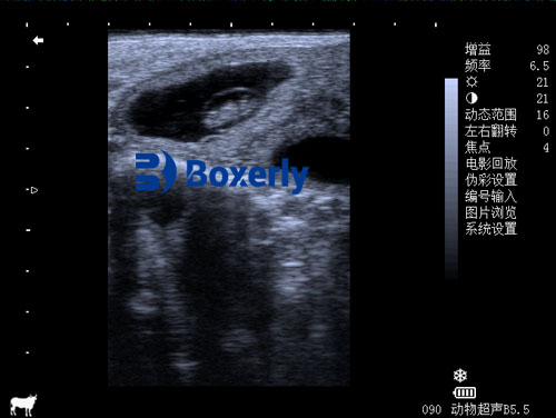
In modern dairy farming, reproductive efficiency is a cornerstone of profitability. Assisted reproductive technologies (ART) such as superovulation (SOV) and embryo transfer are widely used to increase the genetic gain and reproductive output of elite cows. A key to optimizing these protocols is understanding how the cow's ovaries and reproductive tract respond to hormonal stimulation. Among the many tools available, Veterinary ultrasound has emerged as a safe, real-time, and non-invasive technique to monitor the reproductive organs before, during, and after superovulation.

This article explores how ultrasound is used to monitor ovarian changes after superovulation and to assess fetal development in pregnant cows. Drawing on global veterinary practices and published literature, we will examine how this technology helps farmers and reproductive specialists make informed decisions.
Part I: Understanding the Role of the Ovaries in Reproduction
The bovine ovary is a dual-function organ: it releases mature oocytes (eggs) and produces reproductive hormones like estrogen and progesterone. Under normal estrous cycles, a single dominant follicle emerges and ovulates, while the remaining follicles regress. However, with superovulation protocols, cows are administered follicle-stimulating hormones (FSH) to promote the simultaneous development of multiple follicles. This induces the ovulation of multiple eggs, thereby increasing the potential number of embryos available for transfer.
Ultrasound imaging, especially using a rectal probe, plays a central role in evaluating these ovarian responses. In the days following the administration of superovulatory hormones, veterinary ultrasound allows the practitioner to:
Count the number of developing follicles
Measure their diameter
Track ovulation timing
Monitor corpus luteum (CL) formation post-ovulation
Foreign practitioners, especially in North America and Europe, typically perform these evaluations using B-mode (brightness mode) ultrasound devices equipped with linear or convex probes, depending on the size of the animal and the depth required.
Part II: Ovarian Changes Detected by Ultrasound After Superovulation
Under superovulatory treatment (e.g., with FSH followed by a GnRH or LH trigger like PG600 or Receptal), the cow’s ovaries undergo dramatic structural changes. These changes are readily detectable by ultrasound and follow a predictable timeline:
Follicular Growth Phase
Within 2 to 4 days after FSH injections, a cluster of medium to large follicles (ranging from 8 mm to 15 mm in diameter) can be observed on the surface of the ovaries. Unlike a normal cycle where one dominant follicle grows, SOV-treated cows often develop 10 to 25 follicles, each capable of ovulation.
Ultrasound scans during this phase show:
Bilateral ovarian enlargement
Anechoic (dark) circular structures representing fluid-filled follicles
Uniform or mixed sizes depending on the FSH protocol
Ovulation Phase
Following ovulatory stimulation (typically within 24–36 hours after GnRH), the mature follicles begin to collapse and release their oocytes. This is often confirmed via ultrasound by:
Disappearance of previously visualized follicles
Initial signs of corpus hemorrhagicum formation
Slight reduction in ovary size due to follicular rupture
Luteal Phase
Over the next few days, each ovulated follicle transforms into a corpus luteum, which produces progesterone to support pregnancy. On ultrasound, newly formed CLs appear as:
Hypoechoic to isoechoic structures relative to ovarian tissue
Sometimes containing a central cavity (especially in the early luteal stage)
Smaller than CLs observed in natural cycles, especially when formed after superovulation
Studies conducted in Canada and Japan have shown that cows undergoing SOV often develop multiple CLs, each smaller than naturally formed CLs due to the competition among follicles for space and vascularization.
Part III: Importance of Ovarian Monitoring After Superovulation
Why is ultrasound monitoring critical after superovulation?
First, it helps confirm successful ovulation. Not all follicles rupture, and some may become cystic.
Second, it allows for counting the number of CLs to estimate embryo recovery potential.
Third, it aids in identifying abnormalities such as luteinized unruptured follicles or hemorrhagic CLs, which can impact embryo quality and pregnancy rates.
Veterinarians in the U.S. and Europe routinely perform follow-up scans at 5 to 7 days post-insemination to determine the luteal profile. Research suggests a direct correlation between the number of CLs and the expected number of embryos collected during non-surgical flushing.

Part IV: Fetal Development Observation via Ultrasound
Once pregnancy is established, ultrasound becomes an indispensable tool for assessing fetal development. While pregnancy can be detected as early as 26 to 30 days post-insemination, advanced fetal measurements allow for accurate gestational aging and early diagnosis of abnormalities.
Here’s how fetal development is typically observed via ultrasound:
Early Pregnancy (26–45 days)
At 26–30 days, the following can be observed:
Amniotic vesicle: visible as a round, anechoic structure within the uterine horn
Embryo proper: small echogenic focus within the fluid
Fetal heartbeat: visible by 28–30 days using high-resolution probes
By 41–45 days, the embryo resembles a "G" shape on ultrasound. Limb buds, spine, and cranial structures may begin to be visualized.
Mid Pregnancy (46–80 days)
As the fetus grows, its body elongates and becomes more distinguishable:
Limb movements can be observed
Crown-rump length (CRL) is measurable
Placentomes begin forming and can reach up to 5 cm in diameter by 80 days
After this stage, due to the increasing fetal size and depth, it becomes challenging to capture the entire fetus on a single scan. Therefore, veterinarians often focus on specific fetal parts (e.g., head, limbs, heart) and measure their dimensions to estimate fetal age.
Part V: Global Insights and Practices
Globally, the use of veterinary ultrasound in superovulation programs is growing rapidly. According to a 2023 review in the Journal of Reproductive Biotechnology, over 70% of dairy farms in developed countries employ routine ultrasonography in reproductive management.
In countries like Australia and Brazil, where embryo transfer programs are common, veterinarians rely heavily on ultrasound not just for diagnosis but also for decision-making:
In Brazil, CL size and vascularity are used to determine suitability for embryo transfer.
In the U.K., early fetal health scoring using ultrasound helps reduce late embryonic losses.
In the U.S., combining ultrasound with Doppler flow imaging enhances the assessment of luteal function.
Additionally, real-time ultrasound data is being integrated with reproductive software and artificial intelligence tools to optimize breeding schedules and improve success rates.
Conclusion
Veterinary ultrasound has revolutionized how dairy farmers and veterinarians monitor ovarian activity and pregnancy progression, particularly in the context of superovulation. By providing a real-time, non-invasive window into the reproductive tract, it allows precise timing of insemination, embryo recovery, and pregnancy management.
From detecting multiple follicles to measuring fetal dimensions, the power of ultrasound lies in its ability to enhance reproductive efficiency while ensuring animal welfare. As technologies continue to evolve, incorporating advanced ultrasonographic tools into daily herd management will become not just an option but a necessity for progressive dairy operations worldwide.
tags:
Text link:https://www.bxlultrasound.com/ns/817.html


