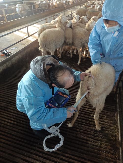
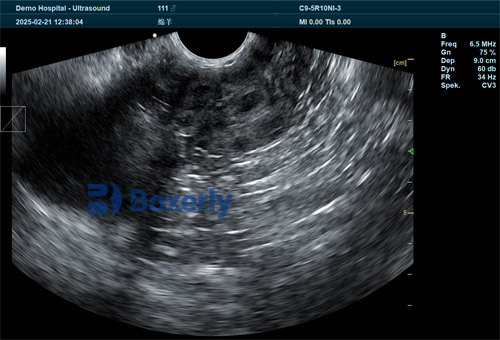
As modern sheep farming evolves, veterinary ultrasonography has become an indispensable tool for reproductive assessment, disease diagnosis, and herd management. Among its most important applications is transrectal scanning, which allows veterinarians and livestock technicians to visualize the internal reproductive structures of ewes with high accuracy. While many farmers are familiar with pregnancy detection using ultrasound, fewer understand the distinct imaging planes—or cross-sections—that can be observed as the ultrasound probe advances through the rectum. These cross-sections reveal a variety of anatomical features critical for evaluating reproductive status, identifying abnormalities, and improving breeding efficiency.

In this article, we explore the three key cross-sections visualized when an ultrasound probe enters the ewe’s rectum during a transrectal examination. By understanding the anatomical landmarks, echo patterns, and practical implications of each plane, sheep farmers and veterinarians can optimize their use of this non-invasive imaging technique. Drawing on global veterinary practices, this article also provides insights from international sheep production systems, where ultrasound use has become routine in both commercial and research settings.
First Cross-Section: The Vaginal and Pelvic Outlet View
When the Veterinary ultrasound probe is first inserted into the ewe’s rectum, the initial cross-section primarily visualizes the area just beneath the rectum—most notably the vagina and the pubic symphysis. This anatomical region marks the beginning of the pelvic canal and provides essential information about the ewe’s reproductive health.
The pubic symphysis appears on ultrasound as a hyperechoic (bright) linear structure with a corresponding acoustic shadow below it, as sound waves cannot penetrate bone. This acoustic shadow is a key reference point in confirming that the probe is appropriately positioned. Beneath the pubic symphysis, the vaginal wall is observed, which—when healthy and free of fluid—displays two hyperechoic serosal layers (the outer peritoneal membranes) with an intermediate hypoechoic muscular and mucosal layer between them. The layered structure, while not sharply distinct due to limited differences in acoustic impedance, can be measured in terms of overall wall thickness.
If vaginal fluid is present, such as during estrus or in cases of infection, a characteristic anechoic (dark) fluid-filled space becomes visible between the mucosal layers, further highlighting the internal vaginal architecture. In addition, large vestibular glands located deep in the vaginal canal may appear as round, hypoechoic regions with scattered echogenic spots, typically measuring about 1.0 × 1.5 cm. These glands are useful indicators of sexual maturity and hormonal activity.
This first cross-section is especially helpful for identifying signs of inflammation, trauma, or retained fluid. Internationally, veterinarians in Europe and North America use this plane to screen for pre-breeding readiness and to identify postpartum complications.
Second Cross-Section: The Cervix and Uterine Body
As the ultrasound probe is gently advanced forward, the second imaging plane comes into view. This cross-section offers a more detailed look at the reproductive tract, specifically the cervix, uterine body, and early segments of the uterine horns. Additionally, the urinary bladder and pelvic inlet can be observed from this angle, providing valuable spatial context.
The bladder appears as a well-defined, pear-shaped anechoic structure, with clear visualization of the bladder wall. Its location—typically anterior to the pubic bone and posterior to the uterine body—helps differentiate urinary versus reproductive organs.
In the ewe, the cervix connects the vaginal vault to the uterine body and is approximately 4 cm long with a wall thickness of about 0.5 cm. On ultrasound, the cervix is identified by its echogenic outer wall and centrally located hypoechoic canal. The uterine body, located just cranial to the cervix, is shorter—around 2 cm in length—and exhibits a wall thickness between 0.3 and 0.5 cm.
One of the most important features of this cross-section is the visualization of the uterine horns. Each horn measures approximately 9 to 12 cm in length and 2 to 3 cm in diameter. They converge at the uterine body and may be slightly asymmetric, often leaning toward the right in multiparous (previously bred) ewes. The uterine horns' echogenic borders and hypoechoic lumen can be easily distinguished in most cases, though horn visibility may depend on parity and reproductive status.
This cross-section is central to reproductive assessment. In the U.S., Australia, and New Zealand, veterinarians commonly use this view during early pregnancy diagnosis or to check for uterine abnormalities such as cysts, adhesions, or pyometra (infection).

Third Cross-Section: Abdominal Reproductive Organs and Digestive Structures
As the probe advances further into the pelvic inlet and enters the abdominal cavity at a 45-degree angle (either left or right), the third ultrasonographic plane reveals a broader field. This includes the uterus, uterine horns, ovaries, and adjacent digestive organs like the rumen and intestines.
This imaging angle is particularly valuable because it allows clinicians to scan for ovarian structures such as follicles and corpora lutea (CLs), which appear as round, hypoechoic or echogenic zones on the ovary. The ovary itself is usually oval-shaped and surrounded by a bright echogenic ring, representing the ovarian capsule and surrounding fat tissue.
In non-pregnant ewes during estrus, small round fluid-filled structures—follicles—are visible, each approximately 0.5 to 1.5 cm in diameter. If the ewe is in the luteal phase, the corpus luteum will appear as a solid, echogenic area, often with central acoustic enhancement. In pregnant ewes, the uterus may contain one or more gestational sacs—anechoic fluid-filled areas housing echogenic fetal structures.
This section also displays portions of the rumen and intestines. The rumen appears as a large hyperechoic wall with posterior shadowing, particularly visible after feeding due to gas content and ingesta. The intestines may exhibit gas-induced reverberation artifacts, resulting in a cloudy echogenic pattern with posterior acoustic shadowing.
In international practice, this third cross-section is indispensable for assessing ovarian cycles, monitoring follicular development for artificial insemination or synchronization programs, and detecting early pregnancy. It is also useful for differentiating reproductive versus gastrointestinal abnormalities in cases of abdominal distension or discomfort.

Clinical Significance and Practical Considerations
Understanding these three cross-sections is not merely an academic exercise. For practitioners in the field—especially those engaged in reproductive management or artificial breeding—this knowledge has practical applications:
Timely and accurate pregnancy detection: Identifying fetal sacs in the third cross-section confirms pregnancy as early as 25 days post-breeding.
Assessment of fertility status: Visualizing the ovaries and uterus allows for estrus synchronization monitoring and evaluation of failed conception.
Early detection of disease: Pyometra, endometritis, or tumors may present as abnormal fluid accumulation or structural irregularities in any of the three planes.
Enhanced training for technicians: As more sheep operations adopt veterinary ultrasonography, training programs in Europe and Australia increasingly emphasize mastery of cross-sectional anatomy.
Conclusion
Veterinary ultrasonography continues to revolutionize reproductive management in sheep by providing a real-time, non-invasive window into the ewe’s internal reproductive system. Understanding the three distinct cross-sections visualized during transrectal scanning—the vaginal and pelvic outlet view, the cervix and uterine body plane, and the abdominal reproductive view—enables more precise diagnoses and improved reproductive outcomes. Globally, veterinarians and producers alike are leveraging this knowledge to boost reproductive efficiency, minimize economic losses, and enhance animal welfare.
As technology improves and ultrasound machines become more portable and affordable, the importance of understanding these anatomical views will only grow. Whether on a high-tech Australian sheep station or a family farm in the U.K., this technique stands as a vital tool for the future of sheep reproduction.
tags:
Text link:https://www.bxlultrasound.com/ns/816.html


