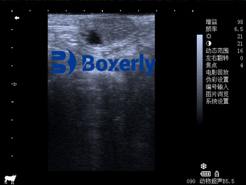
In modern dairy farming, reproductive management plays a vital role in maximizing productivity and profitability. One of the central aspects of reproductive assessment is the identification and evaluation of the corpus luteum (CL), a hormone-secreting structure that forms on the ovary after ovulation. Accurate detection of the corpus luteum can help veterinarians and farmers determine optimal breeding times, detect pregnancy, and diagnose reproductive disorders such as persistent CL. Two main techniques are commonly used for this purpose: rectal palpation and ultrasonography (commonly referred to as ultrasound). While both are widely practiced across the world, each has its strengths and limitations.

This article provides a comprehensive comparison of these two diagnostic methods, with particular attention to their effectiveness at different stages of the corpus luteum's life cycle. We will also include perspectives from international veterinary research and on-farm experiences to help readers make informed decisions on which method to adopt for their herd.
Morphological Stages of the Corpus Luteum
To better understand the differences in diagnostic methods, it is essential to recognize the morphological stages of the corpus luteum. In dairy cows, the CL can be classified into three phases based on days post-ovulation:
Young CL (0–4 days)
Mature CL (5–16 days)
Regressing or Old CL (17–21 days)
These stages are not only important for reproductive timing but also influence the detectability of the CL using different methods. Each phase is characterized by different sizes, shapes, and tissue structures, which affect the accuracy of rectal palpation and ultrasound diagnosis.
Ultrasound: A Precise and Visual Approach
Ultrasonography has become a gold standard in veterinary reproductive imaging due to its non-invasive nature and ability to provide real-time visual feedback. With B-mode (brightness mode) imaging, veterinarians can view internal structures of the ovary and identify the corpus luteum based on its size, shape, and echogenicity.
During the mature phase of the corpus luteum (5–16 days), ultrasound demonstrates high diagnostic accuracy. Studies have shown that ultrasound’s sensitivity (the ability to correctly detect a true CL) is approximately 80.6%, while its positive predictive value (the ability to confirm the CL when detected) is around 85.3%. These values are particularly important in herd-level reproductive management, where minimizing false positives or negatives is critical.

The key advantages of ultrasound include:
Real-time visualization: Allows accurate identification of the CL, follicles, cysts, and uterine contents.
Non-invasive and repeatable: Causes no harm or discomfort to the animal.
Early detection: CL can be seen as early as 1 day after ovulation, appearing as a weak to moderate echoic structure.
Differentiation of CL types: Ultrasound helps distinguish between normal cyclic CL, persistent CL, and pregnancy CL based on echotexture and surrounding ovarian tissue.
Furthermore, the use of Doppler ultrasound (where available) can measure blood flow within the CL, which adds further confirmation of its activity and function.
Rectal Palpation: The Traditional, Tactile Method
Rectal palpation is one of the oldest and most cost-effective methods used in reproductive diagnosis. It involves manually feeling the ovaries through the rectal wall and evaluating the size, shape, and consistency of any palpable structures.
For mature corpus luteum, rectal palpation yields a sensitivity of around 83.3%, which is slightly higher than ultrasound. However, its positive predictive value is lower, at 73.2%, indicating a higher likelihood of false positives. This means that while practitioners may often feel a structure resembling a CL, it might not always be functionally or anatomically accurate.
Rectal palpation is more prone to misdiagnosis during the early (0–4 days) and late (17–21 days) stages of the CL because the structure is either too small, soft, or regressing. Palpation also has limitations in distinguishing between CL and ovarian cysts or other abnormalities, especially in the absence of visual confirmation.
Despite these limitations, rectal palpation remains popular due to:
Low cost: Requires no equipment, only skilled personnel.
Rapid execution: A skilled practitioner can examine many cows in a short time.
Accessibility: Can be performed in remote or under-resourced regions without access to imaging tools.
Diagnosis of Persistent Corpus Luteum
One of the major challenges in reproductive management is identifying a persistent CL, a condition where the corpus luteum fails to regress and continues secreting progesterone, preventing the cow from returning to estrus. This can mimic pregnancy and lead to missed breeding opportunities.
Rectal palpation of a persistent CL may reveal a structure that protrudes from the ovarian surface with a mushroom-like feel and firm consistency. However, such findings are often indistinguishable from a normal pregnancy CL. To confirm persistence, the practitioner typically performs 2–3 palpations over 7–10 days. If the size, shape, and location of the CL remain unchanged, and there is no evidence of pregnancy, the diagnosis is made. In such cases, palpation of the uterus becomes critical, as non-pregnant cows with persistent CL often exhibit mild uterine inflammation or lack of tone.
Ultrasound offers more definitive identification of persistent CL. The echotexture of the structure is typically stronger and more heterogeneous compared to a normal CL. The shape is clearer and the boundaries between the CL and the surrounding tissue are distinct. Ultrasound can also confirm the absence of embryonic or fetal structures in the uterus, providing a definitive diagnosis.
Global Perspectives on Diagnosis Techniques
Veterinary practitioners across the world employ both methods depending on farm size, technology access, and labor costs.
In developed countries such as the U.S., Canada, and much of Europe, ultrasound has largely replaced rectal palpation as the preferred method. It provides faster and more accurate results, especially when used with herd management software that tracks reproductive status, breeding history, and health records.
In developing nations, rectal palpation remains widely used due to its affordability and the shortage of imaging equipment. However, international training programs and technology-sharing initiatives have helped introduce portable ultrasound machines to rural farms, increasing their diagnostic capabilities.
Additionally, studies in countries like Brazil and Australia have shown that combining both methods—initial screening with palpation followed by confirmation with ultrasound—offers a cost-effective yet accurate approach to herd fertility management.
Making the Right Choice on the Farm
When deciding between ultrasound and rectal palpation, farmers and veterinarians must consider:
Stage of the estrous cycle: Ultrasound is better in early detection (0–4 days) and differentiating persistent CLs, while rectal palpation performs well during the mature phase.
Equipment availability: Ultrasound requires an investment in both machines and training.
Accuracy needs: For breeding programs or embryo transfers, ultrasound’s precision may justify the cost.
Labor skill levels: Rectal palpation relies heavily on practitioner experience, while ultrasound reduces the subjectivity of diagnosis.
Conclusion
The accurate detection and evaluation of the corpus luteum are essential for reproductive efficiency in dairy herds. Both ultrasound and rectal palpation offer valuable tools, but each has its optimal use scenarios. Ultrasound stands out for its accuracy, early detection, and ability to differentiate types of CL, while rectal palpation remains a practical and affordable choice for many producers.
For farms that can afford the investment, ultrasonography is the preferred method due to its versatility and diagnostic power. However, a combined approach—leveraging the speed of palpation and the precision of ultrasound—may provide the best balance between cost and accuracy.
As dairy farms continue to modernize and global collaboration in veterinary science grows, the integration of these techniques will only become more seamless, ultimately improving reproductive outcomes and overall herd health.
References
Ginther, O. J. (1992). Reproductive Biology of the Mare and Cow. Equiservices Publishing.
Fricke, P. M., & Lamb, G. C. (2005). Practical use of ultrasonography in reproductive management of dairy cattle. Theriogenology, 64(3), 478–490.
Barros, C. M., et al. (2012). Identification of persistent corpus luteum in dairy cows using ultrasonography and palpation. Brazilian Journal of Veterinary Research.
Sheldon, I. M., et al. (2009). Influence of uterine disease on cyclic activity and fertility in dairy cows. Reproduction in Domestic Animals, 44(Suppl 3), 20–27.
tags:
Text link:https://www.bxlultrasound.com/ns/809.html


