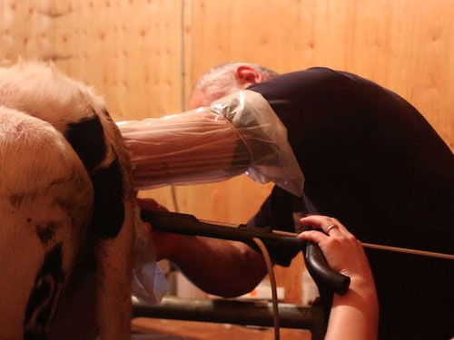
Follicle development in cows is a beautiful, complex dance coordinated by hormones, growth factors, metabolic cues and even ion channels. Most follicles grow normally, one becomes dominant, ovulation happens—and we get another calf. But sometimes something goes awry: a follicle grows, but never ovulates. Instead it hangs out as a cyst, disrupting fertility—and dairy or beef operations can suffer economically.

Let me walk you through what researchers abroad have unraveled about these signals—how follicles develop, and why some become cystic instead of ovulating.
Follicle growth and the hormonal orchestra
From the earliest small follicles through to the dominant preovulatory stage, granulosa and theca cells respond to FSH, LH and estrogen in carefully timed waves. If the LH surge fails or is disrupted, the dominant follicle continues to grow—but never ovulates—and eventually becomes a thin‑walled fluid‑filled structure over ~25 mm, persisting for more than 10 days. That fits the veterinary definition of a follicular cyst in cows.
So what breaks that LH surge? Altered feedback of estrogen on the hypothalamus‑pituitary axis is often blamed. Less Kiss1 neuron stimulation, or changes in GnRH pulse frequency, blunt LH release, and ovulation stalls. On the ovarian side, altered expression of gonadotropin receptors in granulosa or theca cells delays follicle regression and shifts the balance of proliferation versus apoptosis, encouraging cystic persistence.
Growth factors and developmental signals
Folliculogenesis isn’t just hormonal. Inside the follicle, oocyte‑derived signals like GDF‑9 and BMP‑15 guide granulosa proliferation and maturation. In many mammals, GDF‑9 is critical for early follicle progression; its absence stalls development at the primary stage. While the precise interplay in cattle regarding cyst formation isn’t fully mapped, altered GDF‑9/BMP‑15 signaling plausibly disturbs granulosa function, potentially contributing to abnormal follicle persistence.
Another angle: studies of transcriptomes—especially mRNA, lncRNA, miRNA networks—show that in cystic follicles, genes linked to steroid synthesis, energy metabolism, extracellular matrix regulation and steroid hormone receptor signaling are dysregulated. For example, a specific pathway involving lncRNA NONBTAT027373.1, miR‑664b and HSD17B7 has been implicated as a regulator of follicular cyst traits in bovine granulosa cells. That suggests non‑coding RNA networks might underlie altered signaling in follicles that fail to ovulate.
Oxidative stress, apoptosis and autophagy imbalance
One consistent finding: cystic follicles show signs of oxidative stress and increased apoptosis in granulosa cells. Research comparing cows with normal estrus and those with follicular cysts found significantly higher ROS and MDA levels in both serum and follicular fluid, along with lower antioxidant capacity and higher expression of pro‑apoptotic markers (like Bax and cleaved caspase‑3), and lower Bcl‑2.
At the same time, autophagy markers like LC3BII and Beclin‑1 are reduced in granulosa cells from cystic follicles, indicating insufficient protective autophagy. In lab models, a compound like quercetin or an AMPK activator (such as rapamycin) can restore autophagy and reduce apoptosis via SIRT1/AMPK signaling pathways. That raises the possibility that oxidative damage, mitochondrial dysregulation and a failure to clear damaged organelles contribute to cyst formation.
Ion channels, volume regulation and follicle persistence
Here’s a surprising twist from recent Korean cattle data: expression of K₂P potassium channels, especially TASK‑3, is much lower in granulosa cells from large (>25 mm), cystic follicles compared to smaller healthy ones. That goes hand in hand with reduced follicular fluid K⁺ concentration in cystic follicles. Low TASK‑3 expression correlated with signs of cellular senescence: enlarged granulosa cell volume, elevated ROS, calcium and p53/p21 levels, increased SA‑β‑gal staining—all signs that cells have lost normal volume regulation and proliferative control.
Without enough TASK‑3‑mediated K⁺ efflux, fluid dynamics in granulosa cells get disrupted. Cells become senescent, the follicle wall doesn’t regress or luteinize normally, and fluid accumulates—helping sustain the cystic structure.
Stress, metabolism and genetic predisposition
At higher levels—whole animal stress, negative energy balance (NEB), insulin resistance—these also feed into cyst risk. In high‑yield dairy cows early postpartum, NEB and cortisol spikes reduce GnRH/LH, estradiol and LH receptor expression in follicles, delaying ovulation and favoring cyst formation.
Genetic selection for high milk yield also seems to increase cyst incidence: each extra 500 kg yield raises ovarian cyst incidence by about 1.5 %. So a combination of metabolic load, stress, and altered endocrine feedback increases risk.
Putting it all together
Follicular cyst formation in cows appears to involve a convergence of:
| Category | Signals or changes |
|---|---|
| Hormonal disruption | Blunted LH surge, impaired GnRH/Kiss1 feedback |
| Growth factors | Altered GDF‑9 / BMP‑15 signaling, mis‑regulated granulosa function |
| Gene regulation | Dysregulated mRNA/lncRNA/miRNA networks (e.g. NONBTAT027373.1‑miR‑664b‑HSD17B7) |
| Oxidative stress | High ROS, low antioxidants, apoptosis outweighs survival |
| Autophagy imbalance | Reduced LC3BII/Becn1, impaired protective mechanisms |
| Ion channel dysfunction | TASK‑3 downregulated in granulosa cells, leading to senescence |
| Metabolic stress | Cortisol, NEB, insulin resistance, genetic predisposition |
When these signals misfire, ovulation fails and the follicle doesn’t regress normally. Instead it persists, often growing to >25 mm, secreting abnormal estrogen, and failing to luteinize, while suppressing normal cyclicity. Clinically, cows become anestrous or show erratic heats, fertility drops, calving intervals lengthen, and culling risk rises.
Why understanding this matters
Western researchers, particularly in dairy science, emphasize that improving reproductive efficiency means tackling these pathways rather than just treating with hormones. Interventions might include nutritional support to reduce NEB, antioxidant therapy like quercetin, metabolic monitoring, and potentially modulation of ion channel expression or autophagy pathways.
Better genomic selection could reduce predisposition without sacrificing yield. Additionally, monitoring biomarkers like AMH, hormone levels, and expression of key ncRNAs or proteins could allow early detection before a follicle becomes a cyst.
What farmers and vets can take away
Talking transparently with vets or reproduction specialists: when a cow returns to heat irregularly or cycles are absent, consider not just hormone shots but metabolic state, oxidative stress markers, even advanced diagnostics like ultrasonography plus molecular tests. While some markers like TASK‑3 expression aren’t yet practical diagnostics, stress and insulin resistance are manageable through diet and management.
Simple steps can help:
Ensure adequate energy balance after calving
Minimize chronic stress (heat, overcrowding, handling)
Support antioxidant intake (e.g. vitamin E, organic selenium)
Monitor early cycles with ultrasound to detect persistent follicles
In research or high‑value animals, consider more advanced testing of follicular fluid or granulosa cell transcriptomes
Final thoughts
The story of cow follicle development and cyst formation isn’t about one single malfunction—rather a cascade of hormonal, molecular and cellular mis‑steps. Researchers overseas have shown that growth factors like GDF‑9, ion channels such as TASK‑3, ncRNA networks, and the balance between oxidative stress/apoptosis versus autophagy are all players in this drama.
Turning this knowledge into practical farm care means managing stress and metabolism well, supporting antioxidant systems, and working with vets to detect and address follicle persistence before it evolves into chronic cysts. It’s a fine example of how detailed cell‑level and molecular insight pairs with on‑farm practice to support fertility, productivity and animal welfare.
tags: Cow Follicle Development


