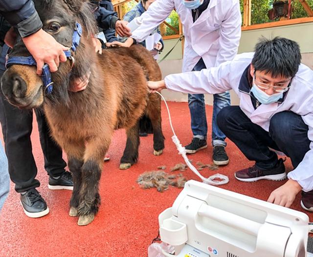
For horse owners and farm vets, keeping mares healthy is not just a job—it’s personal. When something seems off, it can be more than frustrating; it’s worrisome. One of the lesser-talked-about yet serious reproductive issues mares can face is pyometra, a uterine infection that builds up silently but dangerously. Fortunately, ultrasound has become an essential tool to detect this condition early, especially in field conditions where laboratory tests and full veterinary facilities might not be readily available.

Let’s walk through how ultrasound is used to spot pyometra in mares, what signs to watch for, and how real-life farm situations across the world are using this technology to make faster, smarter decisions for their horses’ health.
What Is Pyometra in Mares?
Pyometra is essentially a bacterial infection of the uterus, which leads to accumulation of pus within the uterine cavity. Unlike in dogs, where the cervix often closes completely, mares usually have a partially open cervix, which may allow some discharge. But don't be fooled—many cases are still silent, with no obvious pus draining out. That’s where ultrasound steps in.
Mares suffering from pyometra often have reduced fertility, act lethargic, may show intermittent heat cycles, or none at all. The tricky part is that outward signs are often mild or completely absent, especially in older mares or those not actively bred.
How Ultrasound Helps Detect Pyometra on the Farm
Farm-based ultrasound exams are non-invasive, quick, and safe—ideal for early detection of uterine abnormalities. Portable ultrasound units have advanced significantly over the last decade, with better image resolution, faster scanning speeds, and rugged designs built for barn and pasture work.
Here’s how ultrasound is practically used:
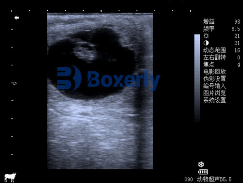
Transrectal Scanning: Most commonly used in mares, where the probe is inserted rectally to visualize the uterus and ovaries. In pyometra cases, the uterus often appears distended, with anechoic (dark) or hypoechoic (grey) fluid filling one or both uterine horns.
Fluid Characteristics: The nature of the fluid can tell you a lot. In simple endometrial fluid (common during estrus), you might see a small amount of anechoic fluid. But in pyometra, you’ll often observe large pockets of cloudy or flocculent material, which suggests pus and infection.
Uterine Wall Changes: Thickened uterine walls, echogenic debris, or visible septa within the fluid can support the diagnosis. Ultrasound can also help differentiate pyometra from uterine tumors, abscesses, or pregnancy.
A Common Scenario on Farms
Let’s say you’ve got a 17-year-old mare that hasn’t taken in two breeding cycles. She’s eating well, no signs of illness. But there’s something “off.” The vet comes out with an ultrasound unit and scans the uterus. On screen, there’s a swollen, fluid-filled uterine horn with cloudy floating particles. That’s classic pyometra.
No bloodwork needed. No guessing. Just a solid, visual diagnosis in under 10 minutes. And that changes everything—because early detection can mean saving the uterus, and sometimes, the mare’s life.

How Vets Around the World Use Ultrasound for Pyometra
In North America, many equine vets rely on routine ultrasound checks as part of pre-breeding exams. If the mare is older or has a history of reproductive issues, checking for uterine fluid is almost standard. In Europe, especially on large breeding farms in Germany and France, veterinarians routinely monitor uterine involution post-foaling using ultrasound—catching potential pyometra cases before they escalate.
Even in developing regions, Portable ultrasound scanners have made it easier to provide high-quality diagnostics in rural or low-resource environments. Some South African vets working in free-range horse communities reported that ultrasound has doubled their early detection rates of uterine infections in mares over the past five years.
Managing Pyometra After Detection
Once pyometra is diagnosed via ultrasound, treatment typically involves:
Uterine Lavage: Flushing the uterus with sterile saline or antiseptic solutions to remove pus.
Antibiotics: Both systemic and intrauterine, depending on the severity.
Hormonal Therapy: Drugs like oxytocin or prostaglandins may be used to stimulate uterine contractions and clearance.
Surgical Options: In rare or severe cases, hysterectomy (uterine removal) may be necessary, though it’s not commonly performed due to cost and risk.
Ultrasound doesn’t just stop at detection—it plays a key role in monitoring treatment progress. Follow-up scans help track fluid clearance, check for recurrence, and confirm the uterus is returning to normal.
Differentiating Pyometra from Other Conditions
One of the biggest benefits of using ultrasound on the farm is its ability to differentiate between look-alike conditions. Not every uterine fluid buildup is pyometra. For example:
Persistent Endometritis: Also presents with uterine fluid, but typically less in volume and with a different appearance.
Pregnancy Loss: Sometimes, a mare that recently lost a pregnancy might retain fluid or tissue that mimics pyometra.
Mucometra: A sterile accumulation of mucus in the uterus—more common in older mares, especially those with cervical damage.
Ultrasound gives you that visual clarity, helping avoid misdiagnosis and guiding the right treatment plan.
Why Early Detection Matters
The earlier pyometra is caught, the better the outcome. In chronic or untreated cases, the infection can:
Scar the uterine lining, reducing future fertility.
Cause systemic illness if bacteria enter the bloodstream.
Lead to colic-like symptoms from uterine distension.
In extreme cases, rupture the uterus, which is life-threatening.
Regular ultrasound checks—especially before breeding—offer peace of mind. For older mares or those with questionable breeding histories, these scans are even more important.
Making Ultrasound a Routine Tool on Horse Farms
More farms are integrating ultrasound into routine care, not just for pregnancy diagnosis, but also for general reproductive health checks. The reasons are clear:
It’s fast: A scan can take under 10 minutes.
It’s portable: Today’s handheld units fit in a vet’s bag or even a coat pocket.
It’s affordable: Costs have dropped significantly, making it accessible for small operations.
It’s preventative: By catching issues early, it reduces long-term costs and improves reproductive success.
One vet in Kentucky summed it up perfectly: “You wouldn’t breed a mare without an ultrasound any more than you’d run a car without checking the oil.”
Closing Thoughts
Pyometra in mares is a hidden threat—but it doesn’t have to be. With proper ultrasound use, early detection is possible, effective, and often life-saving. Whether you're running a high-performance breeding farm or just taking care of a few beloved mares, having access to ultrasound technology is no longer optional—it’s essential.
Seeing the fluid-filled uterus on screen doesn’t just diagnose a problem—it gives you a path forward. That’s what makes all the difference in a mare’s future.
tags:


