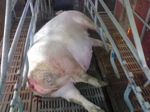
As modern swine production systems continue to evolve toward greater efficiency, animal health and reproductive performance remain critical pillars for profitability. Among the available diagnostic technologies, transabdominal ultrasound has emerged as a key, non-invasive tool in monitoring reproductive status and identifying disorders in sows and gilts. Widely embraced in veterinary practice across North America, Europe, and Asia, this imaging modality allows producers and veterinarians to make more timely and precise decisions regarding breeding management, culling strategies, and overall herd fertility.

In this article, we explore the diagnostic value of transabdominal ultrasound in detecting and managing reproductive disorders in swine. We’ll also examine how international practices, particularly in developed pork-producing regions, have leveraged this technology to improve reproductive efficiency and reduce economic losses caused by undiagnosed infertility.
Understanding Reproductive Disorders in Swine
Reproductive disorders in pigs can be broadly classified into three categories:
Failure to conceive or delayed return to estrus
Pregnancy loss (early embryonic death or abortion)
Anatomical or infectious pathologies of the uterus or ovaries
Common causes of reproductive failure include ovarian cysts, endometritis, silent heat, uterine infections, and poor timing of artificial insemination. If left undiagnosed, these problems can result in extended non-productive days (NPD), increased culling rates, and lower farrowing rates — all of which diminish the profitability of commercial pig operations.
According to a 2022 study from the University of Minnesota’s Swine Health Monitoring Program, reproductive failures are among the top three reasons for sow removal, with more than 25% of culls due to breeding-related issues. This highlights the pressing need for accurate, field-applicable diagnostic techniques.
Role of Transabdominal Ultrasound in Swine Reproduction
Transabdominal ultrasound uses high-frequency sound waves transmitted through a probe placed on the animal’s abdomen to create real-time images of internal structures. In swine reproduction, this imaging is primarily applied between days 18–35 of gestation to confirm pregnancy. However, its utility extends far beyond simple pregnancy diagnosis.
Internationally, particularly in Danish and Dutch swine systems known for their precision farming models, transabdominal ultrasound is widely adopted to:
Detect early embryonic loss or irregular pregnancies
Identify uterine abnormalities such as fluid retention or pyometra
Assess the size and number of developing fetuses
Observe ovarian structures (e.g., cysts or persistent follicles)
Determine whether a sow is truly open or experiencing pseudopregnancy
The use of ultrasound in these contexts is both practical and effective. Because transabdominal scanning is less invasive than transrectal approaches and does not require sedation, it’s well-suited for use on farms with large sow populations. Additionally, ultrasound imaging provides instant visual feedback, allowing veterinarians to make on-the-spot clinical judgments.
Foreign Practices in Reproductive Ultrasound Application
Europe
In countries like the Netherlands and Germany, transabdominal ultrasound is part of standard reproductive protocols. Many farms conduct scans on day 21 post-insemination, followed by a second check on day 35 for confirmation and fetal assessment. High-tech ultrasound units are often integrated with farm management software to automatically update reproductive records, improving traceability and analysis.
Furthermore, some European swine veterinarians have begun utilizing color Doppler ultrasound, which, although less common in field settings, can visualize blood flow in uterine vessels, offering additional diagnostic information for differentiating between viable pregnancies and reproductive pathologies.
North America
In the United States and Canada, transabdominal ultrasound is a staple in breeding herd management. The B-mode imaging technique is the most widely used, where black-and-white images depict cross-sections of uterine contents. Operators are trained to distinguish between anechoic (fluid-filled) gestational sacs, echogenic fetal tissue, and abnormal contents such as pus or gaseous accumulations.
Many U.S. swine operations have integrated portable veterinary ultrasound scanners, which allow real-time scanning directly in gestation barns. These scanners are designed to be rugged, battery-powered, and easy to operate by trained farm staff. Diagnostic criteria and timing protocols are guided by training materials from institutions like Iowa State University and the American Association of Swine Veterinarians (AASV).
Diagnostic Accuracy and Practical Considerations
Several peer-reviewed studies have validated the high accuracy of transabdominal ultrasound in swine reproductive diagnostics. According to a 2021 paper published in Theriogenology, sensitivity and specificity for pregnancy detection using B-mode ultrasound were reported at over 95% when scanning is performed between days 24 and 35 post-breeding.
Accuracy may vary depending on factors such as:
Body condition: Overly fat sows may reduce image clarity due to increased abdominal fat.
Timing: Scans performed too early (e.g., before day 18) are more prone to false negatives.
Operator skill: Experience significantly improves the ability to detect subtle abnormalities.
In addition to biological limitations, there are logistical considerations, such as ensuring proper probe placement and avoiding bladder or intestinal loops that might mimic fluid-filled uterine structures. Training and regular calibration of ultrasound units are essential for optimal performance.

Clinical Scenarios Where Ultrasound Proves Indispensable
1. Distinguishing Between Open and Pseudopregnant Sows
Some sows exhibit signs of pregnancy (e.g., increased appetite, calm behavior) but are in fact pseudopregnant. Ultrasound helps confirm the presence or absence of fetal structures, allowing for timely rebreeding or culling.
2. Diagnosing Pyometra or Uterine Infections
When abnormal uterine content such as hyperechoic fluid or gas patterns is seen, pyometra or metritis is suspected. Early intervention, guided by ultrasound, can save the animal and prevent further herd spread of infection.
3. Monitoring Postpartum Uterine Involution
In high-performing herds, some veterinarians use transabdominal ultrasound to monitor uterine recovery after farrowing, especially in sows with retained placenta or dystocia history. This application is still emerging but shows promising potential.
Future Trends: AI Integration and Enhanced Portability
The future of ultrasound in swine reproduction lies in automation and AI-driven diagnostics. Research from veterinary faculties in Italy and Canada is exploring machine-learning algorithms that interpret ultrasound images and flag potential reproductive issues automatically. This would reduce human error and standardize interpretation across farms.
Moreover, the development of ultra-portable, waterproof units—many with sun-visor screens and extended battery life—makes field use increasingly convenient. Devices such as the BXL-V50 exemplify these advancements, offering high-resolution imaging, rugged durability, and user-friendly design. Though not the focus of this article, such machines are revolutionizing in-barn diagnostics and deserve consideration for modern farms aiming to streamline reproductive management.

Conclusion
Transabdominal ultrasound has transformed how reproductive disorders in swine are monitored and managed. Its real-time, non-invasive capabilities make it an invaluable tool for swine producers and veterinarians worldwide. From early pregnancy checks to complex uterine diagnostics, this imaging modality improves decision-making, enhances sow welfare, and boosts reproductive efficiency.
In an era where every non-productive day costs money and every missed pregnancy impacts farrowing targets, transabdominal ultrasound offers a precise and practical solution. As global pork producers continue to adopt technology-driven approaches, the integration of ultrasound into reproductive health programs will only deepen—fueling better outcomes across the entire swine industry.
References
Knox, R.V. (2021). Use of ultrasound for detecting pregnancy and reproductive issues in swine. Theriogenology, 171, 90–98. https://doi.org/10.1016/j.theriogenology.2021.01.013
Iowa State University. (2023). Swine Reproductive Management with Ultrasound. https://www.vdpam.iastate.edu
American Association of Swine Veterinarians (AASV). (2022). Ultrasound protocols and applications in breeding herds. https://www.aasv.org/library
Wageningen University & Research. (2020). Ultrasound techniques in precision livestock farming. https://www.wur.nl
University of Minnesota Swine Health Monitoring Project. (2022). Culling data reports. https://shmp.umn.edu
tags:
Text link:https://www.bxlultrasound.com/ns/855.html


