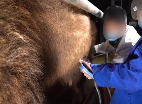
In arid and semi-arid regions where camels play an indispensable role in pastoral livelihoods, the health of these remarkable animals is a major concern for both farmers and veterinarians. As hardy as camels are, they are not immune to gastrointestinal (GI) issues, which can stem from parasitic infections, blockages, diet changes, or systemic diseases. Traditionally, diagnosing intestinal problems in camels relied on physical exams, behavioral observations, fecal analysis, or exploratory surgery. But with the advancement of veterinary imaging technology, one question stands out: Can ultrasound equipment be used to detect intestinal issues in camels?

The short answer is yes—but with some important considerations. In this article, we will explore how ultrasound is applied to camel intestinal health, what foreign experts and researchers have found, and how practical this approach is in real-life field conditions. We’ll also look at why this non-invasive tool is gaining popularity and how it fits into broader camel health management practices.
Understanding Camel Gastrointestinal Problems
Camels have a unique digestive system adapted to dry climates. Like cows, they are foregut fermenters, but they only have three stomach chambers (instead of four), which impacts how feed is broken down and digested. This makes camels vulnerable to a range of GI disorders including:
Impaction (usually due to indigestible materials like plastic or fiber)
Bloat (gas accumulation in the stomach)
Enteritis (inflammation of the intestines, often caused by parasites or bacterial infections)
Colic (abdominal pain with various underlying causes)
Diagnosing these problems early is crucial, especially in working camels used for transport, milk, or meat production. That’s where ultrasound imaging enters the picture.
Ultrasound in Camel Medicine: A Growing Trend
Veterinary ultrasound has become a well-established diagnostic method in cattle, horses, and small ruminants. But for years, its use in camels lagged behind due to the animals’ unique physiology, size, and lack of adapted equipment or training. That has changed recently, particularly in countries like Saudi Arabia, the UAE, Egypt, and parts of East Africa, where camel husbandry is economically and culturally vital.
According to a study published in the Veterinary World Journal (El-Deeb et al., 2020), transabdominal ultrasonography has proven effective in visualizing camel digestive organs such as the intestines, forestomach compartments, and even the peritoneal cavity. This allows for identification of:
Fluid accumulation (suggestive of peritonitis or enteritis)
Gas pockets (indicating bloat)
Masses or blockages (such as bezoars)
Changes in intestinal wall thickness (a marker for inflammation)
These findings confirm that ultrasound is not only applicable to camels—it can provide vital insights into diseases that would otherwise require invasive procedures to diagnose.
How It Works: Ultrasonographic Approach in Camels
Ultrasonography in camels usually involves the B-mode (brightness mode), which gives a two-dimensional image of soft tissues. When examining the abdomen, veterinarians typically use a 3.5 to 5 MHz convex probe for deep penetration through the thick abdominal wall and fat layers.
To perform a scan:
The camel is usually standing and restrained gently to reduce stress.
The right or left flank is shaved or wetted to ensure good contact.
The probe is moved across the abdomen in a systematic pattern.
Real-time images show the structure, movement, and content of intestinal loops.
Abnormalities are identified based on:
Size of the intestinal loops
Motility (peristalsis)
Presence of echogenic (solid) or anechoic (fluid/gas) content
Thickness of intestinal walls
In cases of impaction, the intestines may appear enlarged, immobile, and filled with dense material. Colitis may reveal thickened walls and increased fluid content. Bloat shows up as gas shadows that obscure surrounding structures.
Advantages of Using Ultrasound in Camel GI Diagnosis
Ultrasound offers several advantages over traditional diagnostic methods:
1. Non-invasive and Stress-Free
Ultrasound does not require sedation or surgery. This is particularly useful in camels, which are often stressed by invasive handling.
2. Real-time Diagnosis
Veterinarians can make immediate decisions during the scan, which is critical in emergency cases like acute bloat or intestinal rupture.
3. Field Usability
Modern portable ultrasound units like the BXL-V50, used in livestock worldwide, allow practitioners to bring high-quality imaging directly to the farm or desert environment.
4. Early Detection
Ultrasound can detect subtle changes in the gut before symptoms become severe, allowing for early treatment and better prognosis.
Challenges and Considerations
Despite its benefits, using ultrasound in camels for intestinal issues comes with some challenges:
Camel Anatomy: The thick skin and deep abdominal organs require high-penetration probes and experienced operators.
Training: Few veterinarians are trained specifically in camel ultrasonography, although this is changing through workshops and veterinary courses in the Middle East and Africa.
Cost and Access: While affordable models like the BXL-V50 are now available, access in remote regions is still limited.
International Perspective: What Do Foreign Vets Say?
In regions where camels are less common, such as North America and Europe, interest in camel medicine has grown alongside the popularity of exotic veterinary practice and zoo animal care. Vets trained in large animal medicine are beginning to see camels in private practice, especially in zoos, safari parks, and even hobby farms.
Dr. Jennifer Ketzis, a veterinary parasitologist at Ross University, notes that “Ultrasound has made a big difference in diagnosing gastrointestinal problems in animals that are hard to palpate or scope, like camels. With the right probe and technique, even small intestinal obstructions can be picked up early.”
In Saudi Arabia, Dr. Fahd Al-Mubarak has led several field studies using portable ultrasound devices for diagnosing camel diseases, noting that “This method is safe, effective, and easy to teach camel owners how to interpret the images.”
Case Studies and Practical Outcomes
Case Study 1 – Enteritis in a Lactating Camel (Egypt):
A 5-year-old she-camel presented with diarrhea, weight loss, and lethargy. Ultrasonography revealed increased fluid in the intestines, thickened walls, and sluggish motility. After lab tests confirmed a Clostridium infection, antibiotic therapy and fluid support were initiated, and recovery was achieved in 5 days.
Case Study 2 – Abdominal Mass in a Racing Camel (UAE):
A young racing dromedary displayed colic signs and appetite loss. Ultrasound showed a well-defined echogenic mass in the small intestine. Surgery revealed a bezoar (plant fiber ball), which was removed. The animal returned to racing 6 weeks later.
Conclusion
So, can ultrasound equipment detect intestinal problems in camels? Absolutely. From diagnosing life-threatening impactions to tracking enteric inflammation, ultrasound has become a powerful, non-invasive diagnostic tool for camel gastrointestinal health. While challenges remain in terms of training and accessibility, the benefits—especially in early diagnosis and real-time decision-making—are undeniable.
With more portable and affordable machines like the BXL-V50 now available, the future of camel health monitoring is brighter than ever. For camel owners, veterinarians, and researchers alike, integrating ultrasound into routine care offers a deeper look into the health of one of the world’s most resilient livestock species.
References:
El-Deeb, W. M., Fayez, M., & Elmoslemany, A. (2020). “Diagnostic value of ultrasonography in abdominal disorders of dromedary camels.” Veterinary World, 13(6), 1124–1131. https://www.veterinaryworld.org/Vol.13/June-2020/16.pdf
Mohamed, T., & Hamed, M. (2019). “Use of ultrasonography in camels (Camelus dromedarius): A review.” Open Veterinary Journal, 9(2), 190–197. https://www.ncbi.nlm.nih.gov/pmc/articles/PMC6664207/
Ketzis, J. K. (2021). Interview, Ross University School of Veterinary Medicine, St. Kitts.
Al-Mubarak, F. (2022). Field notes on camel ultrasound training sessions, Riyadh Camel Clinic.
tags:
Text link:https://www.bxlultrasound.com/ns/851.html


