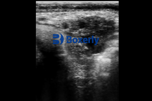
As a cattle veterinarian or livestock producer, precise knowledge of fetal and extrafetal development in pregnant cows is essential for optimizing herd health, improving reproductive efficiency, and making timely management decisions. Ultrasonography—particularly real‑time B‑mode scanning—offers a non‑invasive window into the gravid uterus, enabling visualization and measurement of the gestational sac, fetal membranes, placenta (placentomes), and the developing fetus itself. In this article, we’ll explore how ultrasonography is applied to pregnant cows, describing the characteristic sonographic appearances of key extrafetal and fetal structures throughout bovine gestation and highlighting practical applications for early pregnancy diagnosis, fetal aging, viability assessments, and anomaly detection.

Bovine Gestation Overview
Cows have a gestation length averaging 280–284 days, divided into early (Day 0–90), mid (Day 91–180), and late (Day 181–280) trimesters . Embryonic and fetal development proceeds through predictable stages:
Days 16–18: Formation and extension of placental membranes into both uterine horns.
Day 21–25: Detection of fetal heartbeat under ideal conditions (sometimes as early as Day 21).
Days 30–50: Visualization of placentomes (cotyledon–caruncle complexes), amniotic and allantoic sacs, and early fetal organs.
Days 60–120: Clear delineation of fetal organs—stomach, urinary bladder, spine—and measurement of key biometric parameters for gestational aging.
After Day 120: Assessment of fetal position, viability, and potential for fetal sexing (55–90 days offers highest accuracy).
Ultrasonography Technique in Cows
Transrectal versus Transcutaneous Scanning
Transrectal ultrasonography uses a 5–7.5 MHz linear or sector probe inserted into the rectum, placing the scan head directly against the uterine horns. This approach is ideal for early pregnancy detection (Day 20–60) and detailed fetal imaging.
Transcutaneous (flank) scanning employs a 3.5–5 MHz convex probe placed over the right flank, with the cow standing and restraint minimal. It’s suitable for mid‑ to late‑gestation assessments (Day 60+), twin detection, and placentome evaluation.
Equipment and Settings
Frequency: Higher frequencies (7.5–10 MHz) yield finer resolution for early embryonic structures but have limited penetration; lower frequencies (3.5–5 MHz) are used for deeper structures in larger cows.
Image optimization: Adjust gain, depth, and focal zones to clearly differentiate hypoechoic (dark) fluid‑filled spaces from hyperechoic (bright) fetal tissues and placentomes.
Animal handling: Cows should be calm and secured in a headlock or chute. Remove feces from the rectum and apply coupling gel for optimal probe contact.
Extrafetal Structures
Gestational Sac and Embryonic Vesicle
Days 16–18: The earliest sonographic landmark is the gestational sac, appearing as a small hypoechoic (fluid) chamber within the uterine horn. By Day 18, the lining of the sac becomes hyperechoic relative to the endometrium.
Days 20–25: The embryonic vesicle enlarges to a lemon‑shaped structure; its diameter correlates with gestational age and can be measured to estimate conception date.
Fetal Membranes
Amnion and Allantois: Secondary to the gestational sac, two hyperechoic membranes gradually fuse; the amnion envelops the embryo, while the allantois expands to accommodate growing fetal fluids. Separation of these layers can sometimes be visualized as thin echogenic lines.
Yolk Sac: Transiently visible in early gestation, the yolk sac appears as a tubular hyperechoic structure ventral to the embryo around Day 25–30.
Placentomes (Cotyledon–Caruncle Complexes)
Days 30–40: Placentomes manifest as rounded hyperechoic buttons on the fetal side of the chorion, with corresponding caruncles on the maternal endometrium. The number, size, and echotexture of placentomes provide indicators of placental health and fetal nourishment.
Twin Pregnancies: Transrectal scans can detect two discrete placentome clusters by Day 40–70, enabling early management of twin‑bearing cows to mitigate risks of embryonic loss and dystocia.

Fetal Structures
Embryo and Heartbeat Detection
Day 21–25: Under optimal conditions, the tiny embryo appears as a hyperechoic “dot” within the gestational sac; directed Doppler can confirm cardiac activity. Detection of a rhythmic flicker (“heartbeat”) at this stage confirms viability.
Biometric Measurements for Fetal Aging
Sonographic measurements provide objective estimates of gestational age—critical when breeding records are unknown:
Crown–Rump Length (CRL): Measured along the embryo’s long axis during Days 30–60, correlating linearly with age.
Biparietal Diameter (BPD): Width of the fetal skull at the parietal bones, measured after Day 60.
Thoracic and Abdominal Diameters: Reflect overall body growth; used in regression models to predict age (thoracic diameter P<0.01; abdominal diameter P<0.01).
Femur Length: Evaluated in mid‑gestation (Day 80+), assisting in growth trajectory assessments.
Organ Visualization and Anomaly Detection
Days 60–120: The stomach (anechoic sac), urinary bladder, and portions of the intestinal tract become discernible as anechoic or hypoechoic areas surrounded by echogenic walls.
Skeletal Components: Limb buds, vertebral bodies, and skull bones appear as hyperechoic lines and dots once ossification commences (Days 50–70).
Fetal Sexing: Identification of genital tubercle position allows sex determination between Days 55–90 with high accuracy (>95%).
Clinical Applications of Fetal and Extrafetal Ultrasonography
Early Pregnancy Diagnosis
Detect pregnancy by gestational sac visualization as early as Day 21, but routine practice favors scans at Day 28–35 for reliability and operator ease.
Pregnancy Loss Monitoring
Re‑scan at Days 45 and 60 to confirm ongoing viability; document any embryonic or fetal loss to inform herd reproductive reviews.
Gestational Age Confirmation
Apply biometric formulas to estimate age when breeding records are unavailable, facilitating appropriate nutritional and management plans.
Fetal Viability and Health Assessment
Evaluate heart rate, movement, and amniotic fluid volume to identify at‑risk pregnancies needing intervention.
Fetal Sexing and Twin Management
Plan herd replacement programs and nutritional adjustments based on anticipated calf sex; manage twin pregnancies proactively to reduce complications.
Detection of Anomalies
Early identification of hydramnios, hydroallantois, fetal malformations, or uterine pathologies allows timely culling or treatment, preserving cow welfare and farm economics.

Practical Tips for On‑Farm Scanning
Probe Care: Sanitize with veterinary-approved disinfectants; use a protective sheath for rectal probes.
Operator Training: Proficiency develops with regular practice; consider structured ultrasound courses.
Record Keeping: Photograph key findings and log measurements alongside cow IDs and dates to build individual pregnancy profiles.
Animal Comfort: Use minimal restraint and gentle handling to reduce stress; provide a clean, non‑slippery surface.
Conclusion
Ultrasonography has revolutionized bovine reproductive management by providing real‑time, non‑invasive insights into fetal and extrafetal structures from early gestation through term. Mastery of B‑mode scanning techniques, coupled with knowledge of the sonographic hallmarks of gestational sac formation, fetal membranes, placentomes, and fetal organogenesis, equips veterinarians and producers to make data‑driven decisions that enhance reproductive efficiency, calf outcomes, and overall herd profitability. As ultrasound technology continues to advance—with improved resolution, Doppler capabilities, and portable units—its adoption on cattle operations worldwide will only grow, cementing its role as an indispensable tool in modern livestock management.
References
Early pregnancy diagnosis via ultrasonography allows visualization of fetal heartbeat as early as Day 21 and reliable detection from Day 28 onward. (cdn.ymaws.com, pmc.ncbi.nlm.nih.gov)
Gestational sac and embryonic vesicle development timelines (Days 16–25) in cows. (beefskillathon.tamu.edu)
Biometric associations: thoracic and abdominal diameters correlate significantly with gestational age (Days 73–190). (pubmed.ncbi.nlm.nih.gov)
Placentome visualization and twin detection by transrectal ultrasonography at Days 40–70. (imv-imaging.com)
Fetal organ visualization, viability, and sexing parameters (Days 55–90). (animalrangeextension.montana.edu)
Gestation length in cows (280–284 days).
“Ultrasound Aspects of Fetal and Extrafetal Structures in Pregnant Cats,” adapted conceptually to bovine anatomy and development. https://journals.sagepub.com/doi/10.1053/jfms.2001.0153
tags:
Text link:https://www.bxlultrasound.com/ns/849.html


