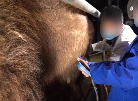
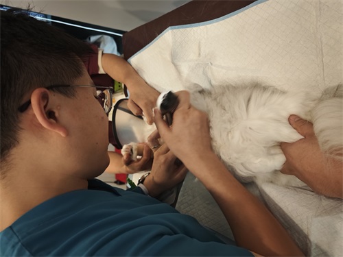
For most veterinarians and animal breeders, ultrasound is one of the most reliable tools in modern animal healthcare—whether you’re checking a dairy cow’s pregnancy status or monitoring muscle development in a finishing pig. But here’s a question that often pops up in real-world veterinary practice: Can a large animal ultrasound machine be used to scan smaller animals like dogs, cats, or even rabbits? It’s a simple question on the surface, but the answer depends on how well you understand both the machine and the animal it’s being used on.

This article unpacks that question from a practical, experience-driven angle. We’ll explore how large animal ultrasound systems work, what matters when scanning smaller bodies, and where the boundaries of this crossover application lie. This isn't a theoretical discussion—many field vets, especially in rural areas or mixed practices, have faced this exact scenario.
Understanding Large Animal Ultrasound
Ultrasound imaging, or sonography, uses high-frequency sound waves to create real-time images of soft tissue structures inside an animal’s body. In livestock, this includes applications like pregnancy detection, fat thickness measurement, or ovarian monitoring. The core principle is the same across species—sound waves go in, bounce off tissue interfaces, and return to form an image. What changes are the machine’s specifications, probe type, and frequency settings.
large animal ultrasound machines are typically designed to accommodate deeper tissue structures and thicker body walls. That means they often use lower-frequency probes (between 2–5 MHz), which offer deeper penetration but lower resolution. This is ideal for scanning a cow’s uterus or a sow’s backfat layer. However, when used on smaller animals—where target structures are much closer to the surface and significantly finer in scale—things get trickier.
What Happens When You Use a Large Animal Ultrasound on Small Species?
Let’s say a vet on a sheep farm is asked to check a farm dog for pregnancy, and the only available equipment is a portable bovine ultrasound. Technically, the machine will still produce an image, but there are limitations.
1. Resolution vs. Penetration Tradeoff
Low-frequency probes penetrate deep, but they lose detail. That means fine structures—like a cat fetus or a small dog’s bladder—might appear fuzzy or be missed entirely. The detail just isn’t sharp enough.
2. Probe Size Matters
Large animal ultrasound probes are physically bigger, and not always ergonomically suited to scanning small or delicate body parts. Trying to image a rabbit’s abdomen with a convex 60mm cattle probe can feel like painting your nails with a broom.
3. Machine Software and Presets
Modern ultrasound machines are often programmed with presets tailored to species—“bovine rectal,” “ovine abdominal,” etc. These influence gain, depth, contrast, and tissue harmonic imaging. When these are optimized for cattle or swine, using the same settings for a cat or dog can compromise image quality. However, some higher-end machines allow for custom presets or manual adjustment, which can significantly improve the outcome.

When Does It Work?
Despite these limitations, there are cases where a large animal ultrasound machine can be adapted to smaller species.
1. Emergency Use in Rural Clinics
In mixed practices, a vet may not have the luxury of a small-animal dedicated scanner. In emergencies—say, checking a small dog for internal bleeding after a fall—a large animal ultrasound can still offer life-saving information, especially if the vet is experienced.
2. Mid-Sized Animals like Goats, Dogs, or Young Pigs
If the subject isn’t tiny and the anatomy isn’t deeply recessed, many mid-range large animal machines can deliver satisfactory images. For example, scanning a goat’s uterus during early gestation with a 5 MHz convex probe can provide clear confirmation of pregnancy.
3. Flexible and Multi-Species Machines
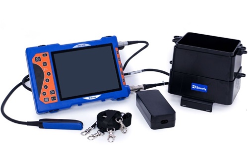
Some portable units, such as the BXL-V50, are built to bridge this gap. These machines come with a variety of probes (including linear ones for shallow imaging), adjustable frequency ranges, and customizable software presets. Vets have reported using such systems for both cow ovary imaging and dog bladder exams with good results.
Real-World Perspectives from Mixed Practice Vets
Dr. Lillian Marsh, a rural vet in Saskatchewan, Canada, regularly switches between cattle, sheep, and small animals in her mobile practice. “I use a portable ultrasound designed for cattle, but I’ve scanned countless farm dogs with it. You just learn how to work around the limitations—turn the gain down, shorten the depth, switch to the linear probe.”
Similarly, in Queensland, Australia, Dr. Tom Reeves uses one unit across everything from Brahman bulls to pregnant border collies. “In my experience, the key isn’t just the machine—it’s knowing what you’re looking for and how to adjust. Sure, I can’t get the same detail as a high-end small animal unit, but I can still make the right call most of the time.”
The Importance of Probe Selection and Settings
Veterinarians working across species must be skilled in adjusting depth, gain, focus, and frequency manually. In the absence of species-specific presets, here are some common tactics:
Switch to a linear probe for higher-resolution shallow imaging, especially useful in small dogs and cats.
Reduce depth to bring superficial organs into view more clearly.
Adjust gain and contrast carefully to reduce “noise” that can blur small structures.
These adjustments may not fully replicate a high-resolution image from a dedicated small-animal ultrasound, but they can make the difference between seeing enough—or not seeing at all.

When Should You Not Use Large Animal Ultrasound on Small Pets?
Despite all these workarounds, there are times when using a large animal scanner on small pets is simply not appropriate:
Cardiac assessments in cats or small dogs, where detailed wall motion and valve function must be seen clearly
Detailed abdominal scans in toy breeds or exotic animals
Neurological or ophthalmic ultrasound, which require high-resolution probes typically operating at 7–15 MHz or more
In such cases, a dedicated small-animal ultrasound is not just ideal—it’s necessary. Using inappropriate equipment risks misdiagnosis, especially in subtle pathologies.
Where the Technologies Are Converging
It’s worth noting that Veterinary ultrasound technology is evolving fast. Manufacturers increasingly build multi-use machines to serve both large and small animals. Compact models now include:
Swappable probes for different depths and applications
Software interfaces with wide frequency modulation
Touchscreen interfaces that allow intuitive adjustments for species size
The BXL-V50, for example, offers veterinary users a high-resolution 8-inch screen, waterproof body (IP56 rated), and compatibility with both convex and linear probes. It’s been used effectively in swine and ruminants but has also been field-tested on dogs and goats—making it an example of how the lines between “large animal” and “small animal” machines are blurring.
Conclusion: One Machine, Many Solutions—With Limits
So, can large animal ultrasound detect small animals?
Yes—with the right conditions. While not a perfect substitute for small-animal units, large animal ultrasound systems can deliver usable images in many small-animal situations, especially in rural or mixed-practice settings. Key factors include the size and condition of the animal, the type of probe used, and the operator’s skill in adjusting the settings.
Veterinary medicine, especially in field settings, is rarely about perfection. It’s about doing what works, when it matters. A capable vet with a flexible ultrasound unit can bridge the species divide when needed. But when image detail matters—such as in cardiac or fine abdominal work—there’s no substitute for the right tool.
Ultimately, ultrasound is less about the machine and more about the mind behind the probe.
References
Whitaker, D. A., & Smith, E. (2021). Veterinary Ultrasonography in Food-Producing Animals. Journal of Veterinary Imaging.
Zwingenberger, A. L., & Wisner, E. R. (2020). Small Animal Diagnostic Ultrasound: Principles and Applications. Elsevier.
Beef Cattle Institute. (2023). Use of Ultrasound for Growth Evaluation in Cattle. https://www.beefcattleinstitute.org/ultrasound-growth
Marsh, L. (2023). Field Notes from a Mixed Practice Vet. Personal Interview.
BXL Veterinary Equipment. (2025). BXL-V50 Ultrasound Scanner Overview. https://www.bxl-vet.com
tags:


