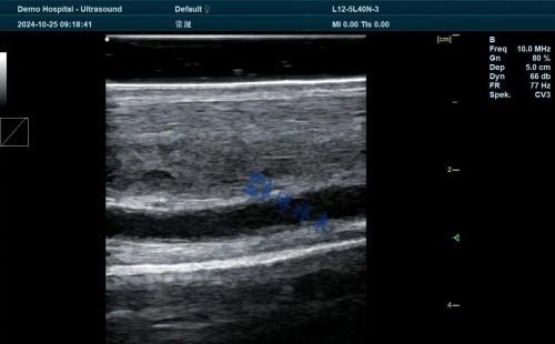
Ensuring the health and well-being of pregnant mares is a top priority for veterinarians, breeders, and equine farm managers alike. In recent years, Doppler ultrasound has emerged as one of the most effective non-invasive technologies for monitoring fetal growth and development in horses. With the rising demand for early pregnancy detection, precise gestational monitoring, and improved foal outcomes, equine Doppler ultrasound is becoming an essential tool in modern breeding operations.

In this article, we explore how Doppler ultrasound is used to monitor fetal growth in pregnant mares, examining key scanning parameters, fetal developmental markers, and insights from veterinary practices abroad. The goal is to provide horse breeders and equine health professionals with a clear, readable, and scientifically grounded guide to using ultrasound technology to enhance reproductive success.
Understanding Equine Pregnancy and the Importance of Monitoring
Pregnancy in mares typically lasts around 340 days, with slight variation depending on breed, climate, and individual health factors. Unlike other livestock, horses exhibit significant variation in fetal growth rate, particularly in mid to late gestation. This makes regular fetal monitoring important not only to assess viability but also to detect abnormalities such as growth retardation, placental insufficiency, or twin pregnancies.
From a practical standpoint, breeders need accurate information to make decisions about nutritional supplementation, exercise, and foaling management. Real-time imaging with Doppler ultrasound provides this information by allowing veterinarians to visualize blood flow, fetal positioning, and key measurements of fetal development.
What Is Doppler Ultrasound and How Does It Work?
Doppler ultrasound uses sound waves to evaluate the flow of blood in vessels. In equine reproduction, color Doppler and pulsed-wave Doppler are most commonly used. These modes detect blood flow velocity and direction, providing insights into both maternal and fetal circulation.
The technology works by emitting high-frequency sound waves through a probe (or transducer) inserted rectally or placed externally on the mare’s abdomen. When these waves encounter moving blood cells, they bounce back with a frequency shift—known as the Doppler effect—which is then interpreted into a visual or auditory signal. This allows veterinarians to “see” fetal heartbeats, placental blood supply, and umbilical cord circulation.

Key Stages for Fetal Growth Monitoring
Early Gestation (14–40 days)
During this phase, Doppler ultrasound can confirm pregnancy, detect multiple embryos (i.e., twins), and evaluate embryonic heartbeat and viability. Around day 25–30, the fetal heartbeat can be detected via color Doppler, offering a powerful confirmation of early pregnancy success. In countries like the U.S. and Australia, early Doppler use has become standard for high-value breeding mares.
Mid Gestation (40–180 days)
At this point, fetal organs begin to develop, and uterine blood flow plays a crucial role in fetal growth. Doppler ultrasound is used to assess the uterine arteries and placental blood vessels. Abnormal patterns—such as decreased velocity or asymmetrical blood flow—can indicate early signs of placental insufficiency, which may result in growth retardation or abortion if not managed.
Late Gestation (180 days to term)
This is the most critical period for fetal growth. The fetus gains most of its weight during this stage. Regular ultrasound assessments help monitor fetal size, position, heart rate, and blood flow in the umbilical artery. In some European equine clinics, veterinarians now measure umbilical artery resistance index (RI) and pulsatility index (PI) to predict potential risks like intrauterine growth restriction (IUGR) or hypoxia.
International Perspectives on Doppler Use in Horses
In the United States, Doppler ultrasound is widely used in Thoroughbred and Quarter Horse breeding programs to reduce pregnancy loss and optimize foaling success. According to a study published by the University of Kentucky’s Gluck Equine Research Center, regular Doppler assessment reduced the incidence of late-term abortion by 17% in high-risk mares.
In Germany, veterinarians often pair Doppler imaging with blood tests to monitor hormone levels like progesterone and estrone sulfate. This combined approach enhances fetal viability assessment, especially in mares with a history of miscarriage or hormone imbalance.
Meanwhile, in the United Kingdom, equine hospitals are increasingly using advanced Doppler imaging equipment to track fetal heart rate variability (FHRV), a promising marker for fetal distress and maturity. Portable machines have allowed field veterinarians to perform these scans on-site, even in remote areas.
Typical Parameters Measured by Doppler Ultrasound
Fetal Heart Rate (FHR)
A steady heart rate between 90–120 bpm is typical in mid to late gestation. Deviations may indicate distress.Umbilical Artery Blood Flow
Low resistance and high velocity are considered normal. High resistance could suggest placental insufficiency.Placental Thickness and Appearance
Increased thickness may be a sign of placentitis. Doppler imaging helps visualize vascular changes linked to infection or inflammation.Amniotic and Allantoic Fluid Volume
While not a Doppler measurement per se, these parameters are often checked during the same session to rule out oligohydramnios or hydroallantois.Uterine Artery Flow
Critical in evaluating maternal blood supply to the placenta. Reduced flow can compromise fetal development.
Practical Benefits for Breeders and Veterinarians
Non-Invasive and Safe: No harm is done to the fetus or mare, and scans can be repeated as needed throughout gestation.
Early Problem Detection: Enables proactive management of at-risk pregnancies, reducing economic losses from foal mortality.
Real-Time Decision Making: Helps decide when to intervene, adjust feed, or plan for assisted foaling.
Portable Equipment: Devices like the BXL-V50 veterinary Doppler ultrasound scanner allow farm vets to scan directly in the stable, increasing accessibility and reducing stress on the mare.
Challenges and Limitations
While Doppler ultrasound provides valuable insights, there are limitations:
Operator Dependency: Image interpretation depends heavily on the experience of the veterinarian.
Gestational Stage Sensitivity: Certain parameters are only meaningful within specific gestational windows.
Cost and Availability: High-quality Doppler machines may be expensive and are not yet common in all rural practices.
However, with growing awareness and training, these barriers are steadily being overcome in both developed and developing equine breeding sectors.

Conclusion
Doppler ultrasound has revolutionized how we monitor fetal growth in pregnant mares. By providing real-time, detailed data on fetal well-being, placental function, and uterine blood flow, this non-invasive technology empowers breeders and veterinarians to make timely decisions that support healthier pregnancies and stronger foals.
As equine reproduction becomes more data-driven, technologies like Doppler ultrasound—especially when used with advanced portable devices—will become standard practice across horse farms worldwide. Whether in Kentucky, Bavaria, or the Australian outback, the ability to “see” fetal development is transforming equine reproductive medicine for the better.
References
Whitwell, K. E., & Smith, E. (2022). Doppler Ultrasonography in Equine Reproduction: Current Practice and Future Directions. Equine Veterinary Journal.
https://beva.onlinelibrary.wiley.com/journal/20423306Gluck Equine Research Center. (2023). Advances in Equine Pregnancy Monitoring Using Color Doppler Ultrasound.
https://gluck.ca.uky.edu/research/doppler-pregnancy-monitoringUK Equine Hospital. (2023). Fetal Heart Rate Monitoring and Umbilical Artery Doppler in Pregnant Mares.
https://www.ukequinehospital.co.uk/services/ultrasound-pregnancyBosh, K. A., Powell, D., & Shelton, B. (2021). Gestational Monitoring in High-Risk Mares Using Doppler Ultrasound. Journal of Equine Science.
https://www.jstage.jst.go.jp/browse/jes
tags:


