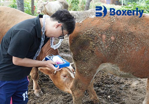
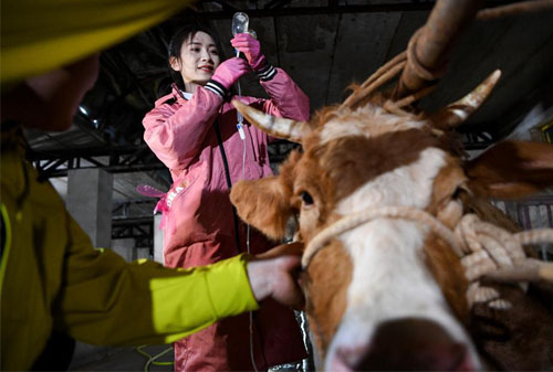
Musculoskeletal injuries—such as tendon tears, ligament sprains, and muscle strains—are common in farm animals like horses and cattle. When an animal becomes lame or swollen, rapid diagnosis is crucial for effective treatment and recovery. In recent years, portable ultrasound imaging has become an indispensable tool for on-site diagnosis. Handheld scanners let veterinarians and livestock managers visualize deep tissue damage immediately, without having to transport the animal to a clinic.

Ultrasound works by emitting high-frequency sound waves through the skin, which bounce off internal structures to create a live image. In the resulting brightness-mode (B-mode) image, normal tissues and fluids appear in characteristic shades of gray and black. Dense structures like bone reflect sound strongly and appear bright, whereas fluid-filled or injured areas reflect less and appear dark. Veterinarians interpret these patterns of echogenicity to distinguish healthy fibers from damage: for example, torn tendon fibers often show a dark (hypoechoic) core on ultrasound.
Advantages of Portable Ultrasound for Injury Diagnosis
Portable ultrasound provides real-time, non-invasive imaging right at the point of care. Unlike X-ray or CT, ultrasound uses no ionizing radiation and can be performed without sedation, causing minimal discomfort to the animal. Devices are now lightweight, battery-operated, and rugged enough for field conditions. The on-site convenience means veterinarians can scan an injured limb or muscle immediately, which greatly cuts down diagnosis time. Key benefits include:
Immediate, on-site diagnosis: Scans can be done in the barn, stable, or even in the pasture. This eliminates delays and risks associated with transporting a lame animal to a clinic.
Non-invasive and safe: Ultrasound causes no pain or harm. Animals often tolerate it calmly, unlike more invasive tests. (Older imaging methods like surgical exploration or even sedation-based MRI carry higher stress).
Real-time feedback: The veterinarian sees live images and can adjust the probe to fully assess the injury. This immediate feedback speeds up decision-making on treatments or rest.
Improved diagnostic accuracy: Ultrasound can reveal subtle soft-tissue damage that is hard to detect by palpation alone. A veterinary review notes that musculoskeletal ultrasonography has become an “established diagnostic method” in livestock care, greatly increasing the likelihood of a definitive diagnosis.
Cost-effective: By reducing the need for expensive referrals (like MRI) and shortening downtime, portable ultrasound often pays for itself. Early injury detection prevents chronic complications and can recoup equipment costs over time.
Guided interventions: Vets can use ultrasound to guide needles and therapies. For example, injecting stem cells or medications into a torn tendon is done under ultrasound guidance to ensure precise placement.
Soft-tissue evaluation: Veterinarians highlight that ultrasound is particularly indicated for examining soft-tissue injuries (tendons, ligaments) that other tests may miss.

Common Injuries and Ultrasound Findings
Farm animals suffer a range of musculoskeletal injuries that benefit from ultrasound assessment. Horses, for instance, frequently incur tendon and ligament injuries during racing or jumping; cattle and sheep may suffer limb trauma from slipping or fighting. Ultrasound helps characterize each injury: some typical examples include:
Tendon tears: A torn superficial digital flexor tendon (common in racehorses) or suspensory ligament will appear as a hypoechoic (dark) region where fiber structure is disrupted. Sonographers scan in both transverse and longitudinal planes: a normal tendon shows fine parallel fibers, whereas an injured tendon appears thickened or has a dark core. Often, even a small core lesion signals a site of fiber rupture.
Muscle and soft-tissue injuries: Injuries that damage muscle (such as a kick, fall, or overextension) create swelling and sometimes fluid accumulation. On ultrasound, a muscle tear may show an irregular fibrillar pattern or localized fluid collection.
Joint and bursa issues: Inflammation inside a joint or tendon sheath often produces excess fluid or synovitis. Ultrasound easily detects these effusions. For instance, a lame cow with hock pain can be evaluated on-farm: ultrasound can show fluid in the joint or tendon sheath, guiding whether to aspirate or inject treatments.
Soft-tissue swellings/abscesses: Lumps or bumps from injury or infection are common. Ultrasound distinguishes a fluid-filled abscess (appearing anechoic or dark on the image) from solid tumors. Veterinarians use this to decide if a swelling needs lancing or can be left alone.
In all these cases, ultrasound gives measurements that tell the recovery story. For example, the cross-sectional area of an injured tendon is often larger than normal, reflecting inflammation. As healing occurs, veterinarians expect to see that tendon thickness decrease and fiber alignment improve. Conversely, a persistent dark area (a core lesion) on follow-up suggests slower healing and a need to extend rest.
Field Workflow and Case Example
In practical terms, portable ultrasound transforms a farm visit. A veterinarian might find a horse with a swollen fetlock. After an initial exam, the vet applies ultrasound gel and scans the tendon region. Within minutes, images reveal whether it is a ligament sprain, tendon tear, or just swelling. For example, one report showed that portable ultrasound identified an early tendon lesion in a racehorse even before obvious lameness occurred. This enabled immediate treatment with rest and anti-inflammatory therapy rather than a missed diagnosis.
Similarly, on a cattle farm a lame bull might receive a quick scan of his hock joint. Ultrasound could show a small effusion or a bursitis, allowing the vet to drain it and bandage the area on the spot instead of sending the animal away for imaging. Handheld devices mean such decisions happen on the spot. GE Healthcare notes that its handheld probes “enable quick assessments of joints, muscles, and tendons, facilitating rapid diagnoses and timely interventions”.
Portable ultrasound also guides treatment procedures. A veterinarian treating a chronic suspensory ligament injury in a horse will use ultrasound to place needles accurately. The device displays the live needle path so that injections of regenerative therapies (stem cells or platelet-rich plasma) hit the precise lesion site. This improves outcomes and avoids blind injections, as emphasized by leading equine sports medicine experts.
Impact on Veterinary Practice
The adoption of portable ultrasound is improving animal welfare and farm productivity. Quick, accurate diagnoses mean injured animals receive appropriate therapy sooner, which can reduce recovery times by weeks or months. By catching problems early, farmers avoid the costs of chronic lameness or severed production (e.g. a dairy cow producing less milk if lame). Veterinarians, in turn, can see more cases per day because exams are faster and more informative. In fact, as portable systems have proliferated, veterinary clinics report that ultrasound has become a routine first-line imaging tool for livestock orthopedics. In the hands of a skilled clinician, a compact ultrasound unit is as valuable as an extra pair of eyes in the field.
Ultimately, portable ultrasound is now seen as a standard part of the large-animal vet’s toolkit. Educational programs and research continue to refine protocols (for example, standard ultrasound views for cattle legs and horse limbs). Industry surveys indicate that most equine and cattle practitioners consider field ultrasound essential for diagnosing soft-tissue injuries and guiding therapy. The evidence is clear: ultrasonography has moved from a specialized niche to a routine practice in large-animal orthopedics.

Conclusion
Portable ultrasound imaging has revolutionized the diagnosis of musculoskeletal injuries in farm animals. By delivering immediate, detailed images of tendons, ligaments, and muscles on the farm, it enables veterinarians to make faster, more accurate diagnoses on site. These timely insights allow treatment to begin sooner, improving recovery outcomes and reducing downtime. In short, handheld ultrasound scanners are now an essential part of modern large-animal practice, helping veterinarians and farmers detect and treat injuries faster while improving animal health and productivity.
References
Leshaw, M. Ultrasonography’s Role in Equine Lameness Cases. The Horse (Mar. 26, 2025). [TheHorse.com].
Kofler, J. Ultrasonography as a diagnostic aid in bovine musculoskeletal disorders. Vet Clin North Am: Food Anim Pract. 25(3):687-731 (2009). DOI: 10.1016/j.cvfa.2009.07.011.
Boxianglai (BXL) Vet. Portable Veterinary ultrasound in Farm – Price and Function. (Dec. 18, 2024). [bxlvet.com].
Boxianglai (BXL) Vet. Veterinary Ultrasound for Horse Tendons. (Dec. 10, 2024). [bxlvet.com].
Chison Medical. Portable ultrasound machine for Veterinary Animals. (accessed 2025). [chison.com].
GE HealthCare. Vscan Air handheld ultrasound systems (MSK imaging). (accessed 2025). [gehealthcare.com].
Black Diamond Equine Clinic. Lameness and Performance. (2025). [blackdiamondvet.com]
tags:


