As a beef cattle professional, mastering the use of ultrasound is essential. It offers a non-invasive, accurate way to assess growth, body composition, and meat quality—particularly backfat, rump fat, ribeye area, and muscle quality—while the animal is still alive. In this article, you’ll learn:
Why and where ultrasound is used in beef production
Detailed preparation techniques, including shaving, cleaning, probe coupling, positioning, and temperature control
Foreign best practices and guidelines (U.S., Europe, international research)
Step-by-step scanning method, referencing the 12th–13th rib and rump areas
Interpreting ultrasound images—what to look for, measurement principles, and common pitfalls
How to use data for feeding, breeding, and marketing decisions
Research findings validating this method
Conclusion and vetted references for deeper insight
1. Why Use Ultrasound in Beef Cattle?
Ultrasound scanning provides live, real-time measurements of key carcass traits without slaughters:
Backfat thickness (BF): shows external fat.
Rump fat (RF): fat at the hip area.
Ribeye area (REA): cross-sectional area of the Longissimus dorsi—indicates muscle volume.
Intramuscular fat (% IMF): marbling within muscle, vital to meat quality.
These traits are moderately heritable and help predict yield, quality, and grade.

2. Foreign Perspectives: Guidelines from Industry
U.S. Extension & Breed Associations (e.g., Mississippi State, Red Angus)
According to Mississippi State:
Backfat & ribfat are measured between the 12th–13th ribs; REA at the same site; rump fat between hooks and pins.
Standard scanning time is ~365 days of age—typical for yearlings.
Red Angus guidelines recommend:
Scanning heifers between 320–460 days with certified technicians.
Clear benchmarking for equipment, clipping, weights, heat, and image submission.
European and Academic Research
Hungarian research (Angus, Simmental, Limousin, Charolais) found:
Ultrasonic REA–carcass REA correlation: 0.74–0.94; average 0.83
P8 fat correlation with carcass EUROP fat score ~0.69.
Meta-analyses show:
Fat thickness accuracy correlation ~.86
Muscle area ~.73
IMF ~.80
with 67% accuracy range: ±0.07″ fat, ±1 sq in REA, ±1% marbling.
3. Preparation: Shaving, Cleaning, Coupling Agents, Temperature
Hair and Debris Removal
Ideal: clip within ½″ and remove dirt .
Alternatives: brush or comb loose hair, but clipped skin is best.
Probe Coupling and Temperature
Apply a coupling gel (or vegetable oil when gel is unavailable; avoid mineral oil) at ~27 °C.
Uniform pressure and probe alignment ensure clearer images.
Equipment Setup
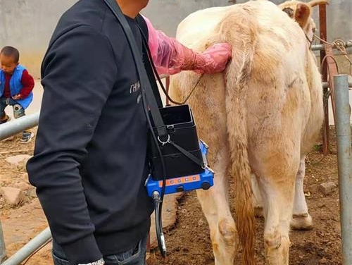
Ensure stable power, shelter, good lighting, and gentle restraint (e.g., chute).

4. Probe Placement: 12th–13th Ribs and Rump
Two key zones require scanning:
12th–13th ribs: for backfat and REA
Rump (hooks to pins): for rump fat
Probe should be parallel to the spine, directly over the rib eye muscle, with the measurement mold pressed evenly.
5. Scanning Technique
Cross-sectional (“transverse”) scan
Measures depth of skin, fat, muscle thickness, and eye muscle area.
Longitudinal (“sagittal”) scan
Evaluates muscle length, fascia, and intramuscular fat organization.
Technician principles:
Correct angles: probe perpendicular to the midline
Optimize image settings: contrast, gain, near-field emphasis
Freeze when image ideal, ensuring clear demarcations of skin, fat, muscle fascia.
6. Image Identification & Measurement
Recognizable Ultrasound Features
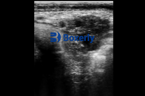
Skin interface: top white line.
Subcutaneous fat: hypoechoic (grey) layer.
Longissimus muscle fascia: bright line below fat.
Ribeye boundary: encircled by fascia.
Measurement Scope
Skin + fat + muscle thickness: via cross-sections.
Longitudinal for intermuscular fat lines, depth assessments.
Use real-time software to measure BF thickness and REA, tagging with animal ID, date, and farm.
7. Data Quality: Errors & Certification
Technician skill greatly impacts accuracy; certification via UGC (or equivalent) recommended.
Avoid angled probe cuts; stay perpendicular.
Clipping and cleaning reduce image artifacts.
Age and weight at scanning must fall within breed-specific windows (e.g., 320–460 days in Red Angus; 365 days standard).
Weight within 7 days of scan ensures accurate EPD adjustments.
8. Using Data: Feeding, Breeding, and Marketing
Feeding & Health Decisions
Monitor growth in real time: decide whether to continue muscle gain, switch to finishing rations (fat accumulation), or prepare for slaughter.
Breeding & Genetic Selection
Use REA, BF, RF, IMF data for EPD calculations. Larger REA = better yield; higher IMF = quality (marbling).
Focus on traits for target markets; e.g., more IMF for markets valuing taste.
Marketing & Sale Timing
Use ultrasound data to choose auction time—maximize market readiness without over-finishing or under-finishing.
9. Research Validation
Studies show ultrasound is a trustworthy proxy for actual carcass analysis:
Hungarian study: REA correlations up to 0.94 in Limousin; overall 0.83 correlation with carcass-referenced REA.
Meta-analysis: fat thickness accuracy ~.86; muscle area ~.73; IMF ~.80.
Other studies: live ultrasound slightly underpredicts yield vs. carcass cut—acceptable for management use.
10. Conclusion
Ultrasound scanning is a powerful, accurate, and animal-friendly method to monitor beef cattle development—from muscle and fat accumulation to carcass quality. The Chinese methodology aligns closely with international best practices: selecting 12th–13th rib scans, removing hair, applying temperature-controlled coupling agents, and ensuring certified techniques.
Benefits include:
Real-time monitoring of growth
Better feeding efficiency and resource utilization
Data-driven breeding and marketing decisions
Non-invasive, repeatable, and welfare-friendly practices
Implementing standardized protocols, staff training, quality control, and certified technicians ensures consistent, trusted measurements. I believe ultrasound will remain essential in modern beef production globally.
References & Additional Reading
Mississippi State Extension: Ultrasound Scanning Beef Cattle for Body Composition –
https://extension.msstate.edu/sites/default/files/publications/publications/p2509_web.pdfRed Angus Association: Ultrasound Guidelines –
https://redangus.org/herd-management/ultrasound/ultrasound-guidelines/Török et al. (2009): Correlation of ultrasonic measured ribeye area and fat thickness… (Hungary) –
https://aab.copernicus.org/articles/52/23/2009/aab-52-23-2009.pdfUltrasound Guidelines Council (UGC): Chapter III – Ultrasonics and the Beef Carcass –
https://ultrasoundguidelinescouncil.org/wp-content/uploads/2021/09/UGC-SG-III-Carcass.pdfIMV Technologies: Use of Ultrasound Scanning Technique–Bovine –
https://www.imv-technologies.com/academy/use-of-ultrasound-scanning-technique-bovineThe Beef Site: Planning a Carcass Ultrasound Session –
https://www.thebeefsite.com/articles/859/planning-a-carcass-ultrasound-session
tags:


