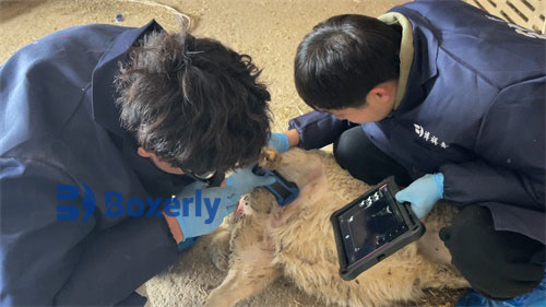
In modern sheep farming, accurate diagnosis and reproductive monitoring are essential for maximizing productivity and ensuring animal welfare. Among the many veterinary tools now available, ultrasound—particularly veterinary B-mode ultrasonography—has become a cornerstone of reproductive assessment. While it is invaluable for early pregnancy diagnosis and fetal development tracking, ultrasound also helps identify developmental abnormalities such as intrauterine growth retardation (IUGR). However, like any imaging technique, ultrasound is prone to misinterpretations due to imaging artifacts, especially when assessing the reproductive organs of ewes.

In this article, we explore how Veterinary ultrasound is used to evaluate fetal development in sheep, with a particular focus on identifying signs of fetal growth restriction. We will also examine the types of imaging artifacts that can occur during the examination of ewe reproductive organs and how to distinguish these artifacts from real anatomical features.
Understanding Intrauterine Growth Retardation in Ewes
Intrauterine growth retardation (IUGR) is a condition where a fetus fails to achieve its expected growth potential. In sheep, IUGR is associated with higher rates of perinatal morbidity and mortality, low birth weight, and impaired neonatal viability. Based on the pattern of fetal growth disturbance, IUGR is typically divided into two types: symmetric (proportional) and asymmetric (disproportional).
Symmetric IUGR: Causes and Ultrasonographic Findings
Symmetric IUGR, also known as proportional growth retardation, occurs when harmful factors affect the fetus during early gestation or even during fertilization. These factors disrupt cell division and organogenesis at a fundamental level. As a result, all fetal dimensions—including head circumference, abdominal circumference, and femur length—are proportionally reduced.
Ultrasound imaging reveals that the fetus is uniformly small. The crown-rump length and head-to-abdomen ratio are within normal limits, but measurements are consistently below gestational age standards. Interestingly, despite the reduced size, the level of organ differentiation often matches the gestational stage. However, the total cell number in these organs is lower than normal, leading to reduced organ mass and function.
Foreign research has linked symmetric IUGR in sheep to a range of causes, including chromosomal abnormalities, congenital viral infections (such as border disease virus), and environmental toxins like radiation or heavy metals. Studies from the University of Adelaide and other leading institutions highlight that symmetrical IUGR often results in high perinatal mortality and low survival rates due to irreversible damage during critical development phases.
Asymmetric IUGR: Causes and Ultrasonographic Features
Asymmetric IUGR typically manifests later in gestation when the fetus has already developed major organ systems. In this condition, head circumference remains relatively preserved while the abdominal circumference is significantly reduced. This results in a disproportionate fetal shape, often described as "brain-sparing" because the fetal body redistributes blood flow toward the brain at the expense of other organs.
Veterinary ultrasound can identify this condition by measuring fetal biometric parameters. The biparietal diameter (BPD) of the head remains appropriate for gestational age, while the abdominal circumference (AC) appears smaller. This discrepancy is a hallmark sign of placental insufficiency, where the placenta fails to deliver adequate nutrients and oxygen to the growing fetus.
Foreign studies, particularly from Spain and the U.S., emphasize that placental insufficiency is often the result of maternal malnutrition, multiple gestations, or chronic hypoxia at high altitude. In sheep farming regions such as the Andes or the Rocky Mountains, veterinarians frequently monitor fetal development in late pregnancy using ultrasound to anticipate complications and intervene early when necessary.

Best Practices for Fetal Growth Assessment in Ewes
Veterinary ultrasound should be performed using a linear or convex transducer with frequencies between 5 and 7.5 MHz for optimal fetal imaging. To ensure diagnostic accuracy, veterinarians and trained technicians often take multiple biometric measurements:
Crown-rump length (CRL) in early gestation
Biparietal diameter (BPD)
Abdominal circumference (AC)
Fetal heart diameter and activity
Placental thickness and echogenicity
Monitoring fetal heart activity also gives clues about fetal stress. In cases of IUGR, heart rates may be reduced, and cardiac dimensions may deviate from normal.
Repeated measurements over time allow the assessment of growth trends rather than relying on a single measurement. When IUGR is suspected, veterinarians may advise dietary supplementation for the ewe or other supportive care to enhance placental function and fetal development.
Distinguishing Ultrasound Artifacts During Reproductive Examination
While ultrasound offers incredible diagnostic power, its accuracy depends on correct image interpretation. Artifacts—false visual patterns that do not represent actual anatomical structures—can lead to misdiagnoses. When examining the reproductive organs of ewes, practitioners should be aware of several common imaging artifacts.
Follicular Artifacts
Ovarian follicles appear as anechoic (dark) circular or oval structures on ultrasound, typically ranging from 3–8 mm in diameter. They are filled with follicular fluid, which does not reflect sound waves, resulting in a black circular image on the screen. However, artifacts may arise when adjacent tissues mimic the echo pattern of a follicle, particularly when scanning from unusual angles.
To distinguish real follicles from artifacts, technicians should perform multi-angle scans and look for consistent shape, location, and size across successive frames. Real follicles also exhibit characteristic wall echoes, and their growth over time can be tracked.
Corpus Luteum (CL) Artifacts
The corpus luteum is a transient endocrine structure formed after ovulation. It typically appears as a hypoechoic (grey) structure with scattered echogenic lines representing vascularization and internal trabeculae. Unlike follicles, the CL has internal structures and does not appear completely anechoic.
Common artifacts include mistaking a collapsed follicle or a hemorrhagic cyst for a corpus luteum. Foreign experts recommend using Doppler ultrasound, when available, to assess blood flow within the suspected CL. Real CLs show robust peripheral blood flow, while cystic artifacts do not.
Corpus Albicans (White Body) Misidentification
The corpus albicans is a regressed form of the CL and may be echogenic due to fibrous tissue. It can resemble a calcified mass or scar tissue on ultrasound. Without proper context, it may be mistaken for a tumor or pathological lesion.
To avoid misinterpretation, it's important to consider the animal’s reproductive history, timing in the estrous cycle, and compare findings with previous scans. Using ultrasound in tandem with hormonal assays (such as progesterone testing) can improve diagnostic accuracy.
Improving Accuracy through Experience and Protocols
One of the key lessons learned from both domestic and international veterinary research is that consistent training and scanning protocols significantly reduce the risk of artifact misinterpretation. For instance, in Germany and Canada, standardized training programs teach technicians to scan from multiple angles, record cine loops, and annotate key findings.
In addition, advances in ultrasound equipment, such as 3D imaging and contrast-enhanced ultrasound, are being explored to improve diagnostic clarity. While still not widely available in sheep practice, these technologies represent the future of precision livestock imaging.
The Importance of Clinical Context
Ultrasound findings should always be interpreted in the context of clinical signs and farm conditions. For example, low fetal measurements accompanied by poor maternal nutrition, high-altitude environment, or concurrent infection may strongly suggest IUGR. On the other hand, a single small measurement in an otherwise healthy pregnancy may reflect measurement error or breed-specific growth patterns.
Conclusion
Veterinary ultrasound has transformed sheep reproductive management by offering real-time, non-invasive insights into fetal health and reproductive anatomy. When used properly, it can detect intrauterine growth retardation, guide interventions, and improve lamb survival rates.
However, practitioners must remain vigilant about the limitations of the technique. Understanding the difference between true anatomical structures and imaging artifacts is essential for accurate diagnosis. Continued training, experience, and technological advances will help ensure that veterinary ultrasound continues to be a reliable and powerful tool for sheep farmers worldwide.
By integrating foreign research, field experience, and practical guidelines, we can better harness the potential of ultrasound in sheep reproduction—improving both animal welfare and farm productivity.
tags:
Text link:https://www.bxlultrasound.com/ns/815.html


