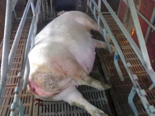
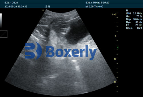
In modern pig production systems, the health and performance of sows and their piglets are critical for achieving high productivity and economic return. One challenge that continues to affect sow reproductive performance and piglet survival is agalactia, or the inability of the sow to produce sufficient milk. Meanwhile, proper management of suckling piglets—including their early nutrition and health—is key to ensuring smooth weaning and optimal growth. Veterinary ultrasound, known for its non-invasive, real-time imaging capabilities, has become an increasingly valuable tool in tackling both of these challenges. This article explores how we use veterinary ultrasound to help prevent sow agalactia and improve piglet management, drawing on international practices and research to explain its growing importance.

Understanding Sow Agalactia and Its Impact
Sow agalactia is a condition characterized by reduced or absent milk production in postpartum sows. It can have a devastating impact on piglet survival, especially during the first 72 hours after birth when piglets are most dependent on colostrum for energy and immunity. Agalactia is often associated with post-partum dysgalactia syndrome (PPDS), a complex of symptoms that includes fever, mastitis (inflammation of the mammary glands), and endometritis (inflammation of the uterus). According to studies from European swine veterinary associations, the prevalence of PPDS in intensive pig farms can reach up to 25%, making early detection and prevention a high priority.
Veterinary Ultrasound as a Preventive Tool in Sows
Veterinary ultrasound, particularly B-mode or real-time ultrasound, allows for the visualization of internal soft tissues, including the uterus and mammary glands. On our farm, and in many others across the globe, we begin using ultrasound on sows around 5–7 days before their expected farrowing date. The goal is to monitor mammary development and detect early signs of mastitis or irregular gland formation.
If any abnormality is detected—such as tissue thickening, fluid accumulation, or glandular asymmetry—intervention begins immediately. Often, this includes adjusting the sow’s diet to ensure proper energy and protein levels, administering preventive antibiotics, or applying anti-inflammatory medications. Internationally, veterinary practitioners from the U.S. and Europe emphasize the importance of combining ultrasound diagnosis with clinical signs such as fever, redness, or pain in the udder to make effective early interventions.

Postpartum Monitoring with Ultrasound
The first 21 days after farrowing are crucial. In our herd management system, we continue to perform ultrasound checks on sows 2–3 times during this period. Postpartum uterine health is a major concern, as lingering infections or poor uterine involution can suppress milk production. By using ultrasound, we assess uterine size, the presence of retained fetal membranes, or fluid buildup, all of which could indicate a developing infection.
If signs of uterine inflammation are visible, we apply targeted treatments such as long-acting oxytetracycline injections or a dose of prostaglandin analogs (e.g., cloprostenol) to promote uterine cleansing and contraction. Literature from Canadian swine health management programs confirms that early uterine treatment significantly reduces the occurrence of PPDS and helps sows return to estrus earlier after weaning.
Promoting Lactation Through Nutritional Support and Imaging
Nutrition plays a critical role in preventing agalactia. We increase energy and protein levels in the sow’s diet after 80 days of gestation, when fetal growth peaks and mammary gland development accelerates. Veterinary ultrasound allows us to track the development of mammary tissue and adjust diets accordingly.
International recommendations from the European Food Safety Authority (EFSA) advise avoiding moldy or mycotoxin-contaminated feed, which can impair immune function and mammary performance. Using ultrasound as a monitoring tool helps us ensure that mammary tissue is developing evenly and that the sow has sufficient body condition to meet the demands of lactation.

Ultrasound's Role in Managing Suckling Piglets
While sow health is paramount, the management of suckling piglets also determines weaning success. One of the critical components in piglet development is early feed supplementation. Beginning as early as 7 days of age, we introduce creep feed—a highly palatable, easy-to-digest solid food designed to supplement sow milk and prepare piglets for weaning.
Veterinary ultrasound can be used to assess the piglet's digestive system, especially in cases where there is poor feed intake or suspected gastrointestinal issues. By scanning the stomach and small intestine, we can evaluate whether digestion is progressing normally or if inflammation, bloating, or blockages are present.
Stimulating Digestive Development
Studies from the Netherlands and Germany have shown that introducing creep feed not only prepares the piglet’s digestive tract for solid food but also stimulates the development of digestive glands and enzyme secretion. Ultrasound allows us to assess the functional development of organs like the pancreas and liver, ensuring the piglets are physiologically ready for the weaning transition.
We start by offering small quantities of feed multiple times per day—typically 4 to 6 times—to encourage frequent intake without overloading the immature digestive tract. Feed remnants are removed before the next feeding to maintain hygiene and prevent spoilage. Monitoring digestive tract structure through ultrasound confirms the effectiveness of this practice.
Ensuring Proper Hydration and Water Access
Access to clean water is vital for piglet digestion and solid feed intake. In our setup, we install nipple drinkers at a height of about 12 cm from the ground, angled downward at 45 degrees. This position aligns with global best practices and encourages natural drinking behavior.
Ultrasound has also been used experimentally in some North American research facilities to measure hydration status in piglets by scanning kidney and bladder size. While not yet a routine practice, this method holds promise for identifying dehydration, which can be especially important during hot weather or in cases of diarrhea.
Practical Benefits of Veterinary Ultrasound in Pig Farming
Veterinary ultrasound brings a suite of advantages to modern pig farming:
Non-invasive and repeatable: Unlike blood tests or surgical interventions, ultrasound does not cause stress or harm to animals. It can be used repeatedly on the same animal to track health status over time.
Early disease detection: In both sows and piglets, ultrasound allows for the identification of issues like mastitis, uterine infections, or gastrointestinal abnormalities before clinical signs become obvious.
Improved decision-making: Data from ultrasound exams help inform treatment decisions, dietary adjustments, and piglet management strategies, improving overall herd health.
Enhanced reproductive outcomes: Regular scanning ensures sows are in optimal condition for farrowing and lactation, while piglets receive early care based on developmental observations.
Global Adoption and Research Insights
Across Europe, North America, and parts of Asia, veterinary ultrasound is becoming standard practice in both small-scale and industrial swine operations. For example, in Denmark—one of the leaders in swine productivity—nearly all breeding sows undergo routine ultrasonographic monitoring. Similarly, in the United States, swine veterinarians are incorporating portable ultrasound devices for in-barn diagnostics as part of precision livestock farming.
Recent research published in journals like the Journal of Swine Health and Production supports the idea that ultrasound can reduce piglet mortality by 15–20% when integrated with other health monitoring tools. The trend is clear: farms that adopt ultrasound-based management protocols consistently achieve better reproductive and economic outcomes.
Integrating Ultrasound Into Daily Management
To incorporate ultrasound effectively, we train our farm staff to use hand-held ultrasound scanners designed for livestock. These devices are user-friendly and provide real-time imaging of reproductive organs, mammary glands, and digestive systems. Once a health concern is spotted, the veterinary team confirms the diagnosis and prescribes the appropriate treatment or nutritional plan.
Routine ultrasound checks are now part of our farm’s standard operating procedures during key stages:
Sow gestation day 80+: Mammary and body condition assessment
5–7 days before farrowing: Mastitis and udder development check
2–3 days postpartum: Uterine and mammary gland imaging
Throughout lactation: Additional imaging if agalactia signs appear
Piglets aged 7–21 days: Digestive tract and hydration scans
Conclusion
In today’s competitive and welfare-focused pig farming environment, preventive care and precise management are more important than ever. Veterinary ultrasound stands out as an indispensable tool in the early detection and management of sow agalactia and in the optimization of suckling piglet development. By allowing real-time, non-invasive monitoring of internal systems, ultrasound supports smarter decisions, healthier animals, and more efficient production.
For farmers worldwide, the integration of ultrasound into daily management routines represents not just a technological upgrade but a transformational shift toward data-driven animal care. Whether you're raising pigs in Europe, North America, or Asia, the message is clear: investing in veterinary ultrasound is an investment in your farm’s future.
tags:
Text link:https://www.bxlultrasound.com/ns/813.html


