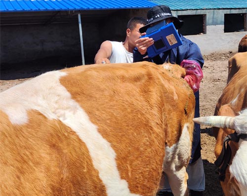
As a livestock farmer, I rely on practical tools to keep my herd healthy and productive. One of the most powerful yet underutilized tools in our diagnostic arsenal is veterinary ultrasonography, especially when it comes to detecting internal conditions like bovine cholelithiasis, commonly known as gallstones. While this condition is relatively rare in cattle compared to other species, it can lead to significant liver and digestive issues if not diagnosed and managed early.

This article explains how ultrasound works in detecting gallstones in cattle, what signs to look out for, and why it is a vital technique for maintaining herd health. I’ll share this from a farmer’s perspective, supported by sound veterinary principles and the latest imaging technologies used in the field.
Understanding Bovine Gallstones
Gallstones in cattle are concretions of bile components, primarily cholesterol, calcium bilirubinate, or mixed substances, that form in the gallbladder or bile ducts. In cattle, these stones are not always associated with clinical signs, but in some cases, they can lead to biliary obstruction, cholangitis, or secondary liver damage.
As a farmer, it’s crucial to know the early signs: decreased appetite, weight loss, intermittent fever, jaundice, abdominal pain, or changes in manure consistency. However, these signs are often vague and non-specific, making it difficult to identify the problem without imaging tools like ultrasound.
What Is Ultrasound and How Does It Work?
Veterinary ultrasound, or ultrasonography, is a non-invasive imaging technique that uses high-frequency sound waves—typically between 1.5 to 18 MHz—to create real-time images of internal organs. These waves are emitted by a transducer, which is placed on the animal’s skin with the help of a transmission gel that improves sound conduction.
As the sound waves penetrate the body, they are reflected back by different tissues. The returning echoes are captured by the transducer, converted into electrical signals, and processed by the ultrasound machine to form an image. B-mode grayscale imaging is most commonly used, which gives a detailed 2D view of internal structures like the liver and gallbladder.
Why Use Ultrasound for Detecting Gallstones in Cattle?
In most farm environments, diagnosing internal liver or gallbladder issues used to be based on clinical symptoms, blood tests, or exploratory surgery. But now, with ultrasound, I can work with veterinarians to visually inspect the gallbladder and surrounding structures in real-time without invasive procedures.
When detecting gallstones:
Gallstones appear as hyperechoic (bright) spots with strong acoustic shadowing on the ultrasound screen.
The gallbladder may be enlarged or distended.
Bile ducts may appear thickened or dilated if there is an obstruction.
In some cases, echogenic sludge in the bile may also indicate early cholelithiasis.
These visual signs, combined with blood work showing elevated liver enzymes, can give us a solid diagnosis without guessing.
Practical Steps on the Farm
When we suspect a gallbladder issue in a cow, here’s the basic process followed during an ultrasound exam:
Preparation: The right side of the cow’s abdomen, particularly the right 9th–12th intercostal spaces, is clipped and cleaned. This is where the liver and gallbladder are best accessed.
Probe Selection: For adult cattle, a low-frequency convex probe (3.5–5 MHz) is used to get deeper penetration. Higher-frequency probes give more detail but are limited in depth.
Imaging: The vet moves the transducer slowly over the liver area. The gallbladder typically appears as a round to oval anechoic (black) structure.
Interpretation: Any echogenic masses within the gallbladder lumen, especially if they cause shadowing, raise suspicion for gallstones. Changes in gallbladder wall thickness or bile duct dilation support the diagnosis.
Documentation: Images are saved digitally (usually in DICOM format) for comparison or referral to specialists if needed.
Limitations and Challenges
Of course, ultrasound has its limitations. Gas in the intestines, a common issue in ruminants, can interfere with the clarity of the image. The deep location of the gallbladder and overlapping structures may make it hard to get a perfect view, especially in obese or heavily muscled cattle.
Also, gallstones may not always be visible if they are small or not calcified. In such cases, indirect signs like gallbladder wall thickening or bile duct abnormalities become critical for diagnosis.
That’s why the success of ultrasonography heavily depends on the experience of the operator and their knowledge of bovine anatomy.
Benefits Over Other Imaging Methods
Compared to radiography (X-rays), ultrasound offers:
Real-time imaging of soft tissues, which X-rays can’t capture effectively.
No exposure to radiation, making it safe for repeated use in pregnant or breeding stock.
Portability, which means we can perform it right in the barn or field.
Guidance for procedures, such as targeted biopsies or aspirations.
These features make it an ideal tool not only for gallstone detection but also for checking reproductive organs, the liver, kidneys, or intestines.
What to Do After Detection?
If gallstones are confirmed, the next steps usually involve:
Supportive care: Including hydration, anti-inflammatory drugs, and dietary management to reduce bile stasis.
Surgery: In rare severe cases, cholecystectomy (removal of the gallbladder) may be considered, though this is complex and not commonly performed in cattle.
Monitoring: Follow-up ultrasounds to track changes in gallbladder size and content.
Most mild cases can be managed medically with close observation, especially if the cow shows signs of recovery and continued weight gain.
Final Thoughts from the Farm
Ultrasound has transformed how we manage herd health on our farm. For conditions like gallstones—which are often silent but potentially serious—early detection through ultrasonography can save animals from severe complications and unnecessary suffering.
It also gives us peace of mind. Knowing we have the tools and knowledge to detect these internal issues means we can intervene early, improve outcomes, and maintain the productivity of our herd.
For any fellow farmers or veterinarians considering integrating ultrasonography into their cattle health program, I highly recommend it—not just for reproduction and musculoskeletal assessments, but also for detecting internal organ issues like gallstones. With the right training and practice, it’s one of the most powerful diagnostic tools in modern veterinary medicine.
tags:
Text link:https://www.bxlultrasound.com/ns/806.html


