Ultrasound imaging, also known as ultrasonography, is a non-invasive, safe, and effective diagnostic tool widely used in modern veterinary practice. In bovine medicine, it plays a crucial role in reproductive management, disease diagnosis, and herd health monitoring. This article provides a detailed overview of how veterinarians perform ultrasound examinations on cows, with a focus on reproductive assessments, common diagnostic uses, procedures, and equipment.

Understanding Veterinary ultrasound
Veterinary ultrasound uses high-frequency sound waves, typically between 2 and 10 MHz, to create images of internal organs and tissues. Unlike X-rays, ultrasound does not use ionizing radiation, making it safe for use in pregnant animals and repeat evaluations.
The sound waves are transmitted from a probe, or transducer, which is placed on the cow’s body. These waves bounce off internal structures, and the returning echoes are processed into real-time images. In cows, this technology is most commonly used transrectally for reproductive examinations, but it can also be applied transabdominally or externally, depending on the target organ.
Primary Uses of Ultrasound in Cattle
1. Pregnancy Diagnosis
The most common reason for ultrasound use in cows is to detect pregnancy. Transrectal ultrasonography allows veterinarians to detect pregnancies as early as 26–30 days post-insemination. This helps farmers make early decisions regarding culling, rebreeding, or managing valuable breeding stock.
Veterinarians assess the size of the fetus, fetal heartbeat, presence of twins, and sometimes even the sex of the calf depending on the stage of pregnancy.
2. Reproductive Tract Evaluation
Besides pregnancy checks, ultrasound is vital for evaluating the uterus and ovaries. Veterinarians can diagnose uterine infections, fluid accumulation, ovarian cysts, and stages of the estrous cycle. This information helps optimize breeding timing and improve fertility rates.
3. Fetal Sexing
At around 55 to 70 days of gestation, experienced veterinarians can determine the sex of the fetus using ultrasound. This is beneficial in managing breeding programs, especially in dairy operations where replacement heifers are highly valuable.
4. Pathology Detection
Ultrasound can be used to detect abscesses, tumors, hernias, liver problems, and intestinal blockages. Though less commonly used in this way for cattle, it is becoming more prevalent with portable, rugged ultrasound units designed for field use.

Equipment and Setup
ultrasound machine
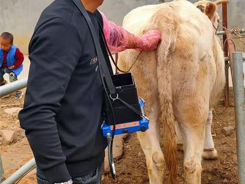
Portable veterinary ultrasound machines are preferred on farms. They typically have a probe (transducer), a display monitor, and a rechargeable battery. Some newer models come with wearable monitors or goggles, allowing hands-free operation during rectal examinations.
Probes
Linear-array transducers are the most common for bovine reproductive ultrasound, typically in the 5–7.5 MHz range. Higher-frequency probes provide better image resolution but less penetration, while lower-frequency probes allow deeper scanning but with lower resolution.
Coupling Gel
An ultrasound gel or lubricant is used to ensure good contact between the transducer and the animal’s rectal wall, reducing air interference and improving image clarity.
Step-by-Step Procedure of a Bovine Reproductive Ultrasound
Restrain the Animal
The cow is safely restrained in a chute to minimize movement and ensure the safety of both the animal and veterinarian.Preparation
The veterinarian dons a long rectal glove, applies generous ultrasound gel to the arm and probe, and gently inserts the hand and transducer into the rectum.Locating the Reproductive Organs
Using palpation and image guidance, the veterinarian locates the uterus and ovaries. The transducer is manipulated to obtain cross-sectional and longitudinal views.Image Interpretation
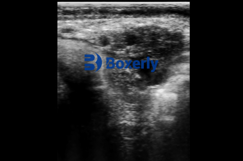
The screen displays real-time images. The veterinarian assesses the uterus for signs of pregnancy, infection, or abnormal fluid. The ovaries are checked for follicles or cysts.Recording and Reporting
Some machines allow image or video capture. The findings are documented, and if necessary, images are shared with other veterinary professionals or stored for later comparison.

Advantages of Ultrasonography in Cattle Practice
Early and Accurate Pregnancy Detection
Compared to manual palpation, ultrasound allows earlier and more precise identification of pregnancy, reducing open days and improving herd productivity.Non-Invasive
No sedation or surgery is required, and the procedure causes minimal stress to the animal.Improved Reproductive Performance
Identifying ovarian activity and uterine conditions helps tailor breeding programs, leading to better conception rates.Enhanced Disease Diagnosis
Ultrasound helps detect internal abnormalities that would otherwise go unnoticed until clinical symptoms appear.
Limitations and Considerations
Operator Skill
Accurate interpretation of ultrasound images requires experience. Misinterpretation can lead to incorrect diagnosis or missed pregnancies.Equipment Cost
High-quality ultrasound machines can be expensive, which may be a barrier for small-scale farms without shared veterinary resources.Limited by Gas and Bone
As with all ultrasound, air (as in intestines) and bone (like the pelvis) can obstruct the sound waves, limiting imaging in certain areas.
New Technologies in Bovine Ultrasonography
Technological advancements have brought about 3D imaging, Doppler ultrasound, and even AI-assisted interpretation. Doppler ultrasound, for instance, is now being explored in cattle to assess blood flow in the uterus and fetal vessels, which could enhance early detection of at-risk pregnancies.
Wireless ultrasound probes and cloud-connected systems are also becoming more common, allowing for remote consultations and telemedicine applications in veterinary practice.
Conclusion
Ultrasound imaging has revolutionized cattle health management, especially in the field of reproduction. Veterinarians skilled in bovine ultrasonography provide invaluable insights that improve breeding outcomes, detect disease early, and enhance the overall productivity of livestock operations. As technology continues to evolve, ultrasound is poised to become even more accessible and powerful for cattle producers worldwide.
References
American College of Veterinary Radiology – Ultrasound Guidelines
https://www.acvr.orgMerck Veterinary Manual – Bovine Reproductive Ultrasound
https://www.merckvetmanual.com/management-and-nutrition/management-of-reproduction-cattle/use-of-ultrasound-in-cattleUniversity of Wisconsin-Madison, School of Veterinary Medicine – Ultrasound in Bovine Practice
https://vetmed.wisc.edu
tags:
Text link:https://www.bxlultrasound.com/ns/802.html


