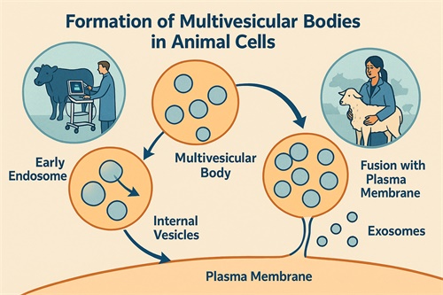
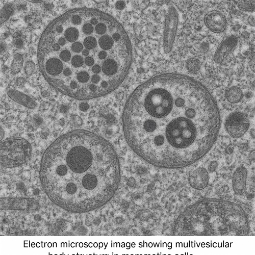
In veterinary medicine, the abbreviation "MVB" typically refers to multivesicular bodies—specialized endosomal structures found in the cells of both animals and humans. These organelles are small vesicles that form inside larger membrane-bound compartments and play a key role in intercellular communication, immune responses, and disease pathology. While MVB may sound like a niche cellular term, its relevance to veterinary diagnostics, particularly in imaging and infectious disease management, is increasing rapidly.
In the context of veterinary practice, especially with the growing application of advanced diagnostic tools such as ultrasound and molecular imaging, understanding the role of MVBs can offer insights into various disease processes in farm animals and companion animals alike.
What Are Multivesicular Bodies?
Multivesicular bodies are a type of late endosome characterized by the presence of multiple small vesicles enclosed within a larger vesicle. These internal vesicles are formed when parts of the endosomal membrane invaginate and pinch off into the lumen of the endosome. Eventually, MVBs can fuse with lysosomes for degradation of their contents, or with the plasma membrane to release extracellular vesicles known as exosomes.

Why Are MVBs Important in Veterinary Science?
Role in Disease Mechanisms
MVBs are involved in the cellular trafficking and disposal of proteins, lipids, and signaling molecules. In animals, these vesicles help manage cellular waste, but more importantly, they play a role in modulating the immune system and in the pathogenesis of viral infections.
In livestock, some viruses—such as foot-and-mouth disease virus (FMDV) and African swine fever virus (ASFV)—hijack the host’s endosomal machinery, including MVBs, to facilitate viral replication or immune evasion. By understanding this process, veterinarians and researchers can better design vaccines or antiviral strategies.
Marker of Cell Health and Cancer Diagnosis
Changes in the number, morphology, and function of MVBs have been observed in various types of cancers, including those that affect companion animals such as dogs and cats. Because they release exosomes into bodily fluids, MVBs can indirectly reflect the state of cellular health. Non-invasive diagnostic tools such as ultrasound-guided fine needle aspiration (FNA) can help obtain samples where such markers may be present.Applications in Diagnostic Imaging
Ultrasound is one of the most commonly used imaging techniques in veterinary practice. While MVBs themselves are not visible via ultrasound, their role in disease processes such as tumors, infections, and inflammation can indirectly be inferred through imaging results combined with cytological or molecular analysis.
For example, in reproductive or mammary gland tumors in dairy cattle or canines, abnormalities that may relate to MVB-driven exosomal signaling can affect the progression or recurrence of tumors. Identifying changes in fluid composition and cellular response through guided biopsy and fluid aspiration, aided by ultrasound, enhances diagnostic accuracy.
Exosome Research in Veterinary Medicine
MVBs are the parent structures of exosomes, and research into exosomes is opening new diagnostic and therapeutic frontiers in veterinary science. Exosomes carry proteins, RNA, and microRNA from the cells they originated from, making them potential biomarkers for diseases like bovine respiratory disease (BRD) and canine lymphoma.
This has sparked interest in “liquid biopsy” methods—non-invasive tests that detect exosomes in blood, milk, or urine samples. Although still developing, this field is quickly evolving and holds promise for more effective disease monitoring in herds and pets alike.

Therapeutic Potential
Beyond diagnostics, MVBs and the exosomes they release are being explored as vehicles for delivering therapeutics in veterinary medicine. Studies have begun to look at the possibility of engineering exosomes loaded with drugs or RNA therapies to target specific diseases in animals—especially chronic inflammatory diseases or cancers.
Conclusion
MVBs may appear to be microscopic curiosities, but their implications in veterinary medicine are wide-ranging. From being markers of disease to potential therapeutic tools, their role is growing as molecular diagnostics and imaging technologies advance in animal health care.
For veterinarians working with livestock, poultry, and companion animals, keeping up with developments in cellular biology—like the role of multivesicular bodies—can lead to earlier diagnosis, better treatment outcomes, and improved overall animal welfare.
References
Kowal, J., Tkach, M., & Théry, C. (2014). Biogenesis and secretion of exosomes. Current Opinion in Cell Biology. https://doi.org/10.1016/j.ceb.2014.01.003
Théry, C., Witwer, K. W., & Aikawa, E., et al. (2018). Minimal information for studies of extracellular vesicles 2018 (MISEV2018). Journal of Extracellular Vesicles. https://www.ncbi.nlm.nih.gov/pmc/articles/PMC6322352/
van Niel, G., D’Angelo, G., & Raposo, G. (2018). Shedding light on the cell biology of extracellular vesicles. Nature Reviews Molecular Cell Biology. https://www.nature.com/articles/s41580-018-0002-6
Veziroglu, E. M., & Mias, G. I. (2020). Characterizing extracellular vesicles and their diverse RNA contents. Frontiers in Genetics. https://www.frontiersin.org/articles/10.3389/fgene.2020.00700/full
tags:
Text link:https://www.bxlultrasound.com/ns/797.html


